Skip to content
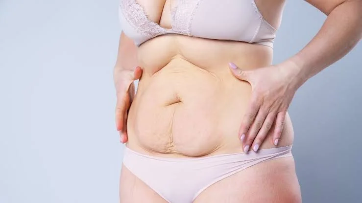
Liposuction is one of the most performed cosmetic procedures worldwide, offering dramatic improvements in body contours by extracting targeted subcutaneous fat deposits. While modern techniques like Vaser and power-assisted liposuction have significantly enhanced precision and reduced recovery times, the body’s natural response to surgical tissue trauma remains an essential part of the healing journey. One of the most common early post-operative complications encountered by patients is fluid accumulation, clinically known as a seroma.
Understanding seroma after liposuction is vital for an uncomplicated recovery. Patients frequently ask what is seroma after liposuction, how to identify its physical signs, what causes seroma after liposuction, and what clinical steps are needed for effective seroma after liposuction treatment. This comprehensive medical guide details the biological mechanisms behind fluid collection, explains how to treat seroma after liposuction, outlines what is the fastest way to heal a seroma, and provides actionable advice to protect your final aesthetic results.
Table of Contents
What Is Seroma After Liposuction and What Causes It?
To understand why this issue develops, we must examine the surgical mechanics: what is seroma after liposuction and what causes seroma after liposuction from a tissue dynamic perspective?
Get Free Consultation
Have any questions? Leave your details and we'll get back to you shortly.
During seroma after liposuction surgery, micro-cannulas travel through the subcutaneous fat layer, mechanically disrupting or liquefying fat cells before extracting them. This process creates a honeycomb-like network of empty space beneath the skin, referred to as “dead space.” Additionally, the physical movement of the cannula damages microscopic lymphatic channels and blood capillaries that normally drain tissue fluids.
Driven by the acute inflammatory response, blood plasma and clear lymphatic fluid leak into these newly created empty cavities. When the volume of fluid produced exceeds the body’s natural reabsorption rate, the liquid pools together, forming a localized fluid pocket known as a seroma.
Key Factors That Increase Seroma Risk:
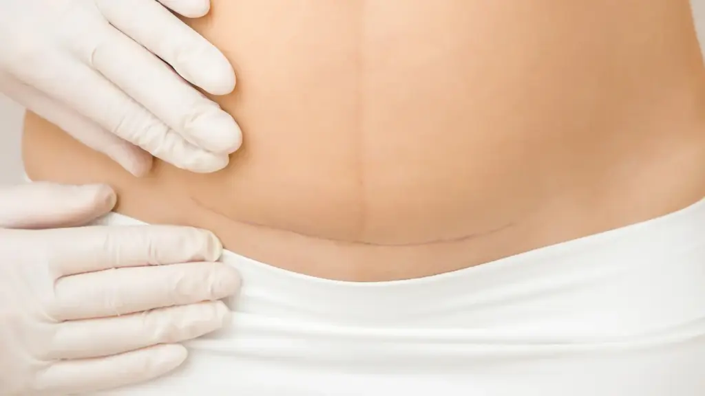
- Large-Volume Fat Extractions: Harvesting extensive volumes creates larger areas of dead space.
- Use of Ultrasound-Assisted (Vaser) Lipo: Thermal energy can cause friction and temporary thermal trauma to surrounding tissues if aggressive settings are used.
- Inadequate Post-Op Compression: Failing to wear prescribed compression garments allows fluid to collect freely in untreated dead spaces.
- Excessive Physical Activity Too Early: Early vigorous movement increases local blood flow and friction between tissue layers, driving extra fluid exudation.
What Does Seroma Look Like After Liposuction?
Recognizing the physical manifestation of fluid buildup allows for timely intervention. What does seroma look like after liposuction? It typically appears as a localized, soft swelling or bulge that behaves differently than general post-operative inflammation:
- Fluctuant Soft Swelling: Unlike hard, fibrotic tissue, a seroma feels soft, squishy, or wave-like when gently pressed similar to a water balloon under the skin.
- Visible Asymmetry or Bulging: A localized protrusion appears in areas like the lower abdomen, flanks, or inner thighs where fat was heavily extracted.
- Clear or Pale Straw-Colored Fluid: If drained, the liquid is thin, translucent, pale yellow, or slightly pinkish (serosanguinous).
- Sensation of Liquid Movement: Patients often report feeling liquid sloshing or shifting beneath their skin when changing positions or walking.
- Mild Discomfort or Tightness: While small seromas are mostly painless, large fluid collections create pressure, tightness, or mild aching due to skin stretching.
How to Treat Seroma After Liposuction: Clinical & At-Home Protocols
Addressing fluid pockets requires a structured approach depending on the volume of accumulated fluid. Here is a detailed breakdown of seroma after liposuction treatment options:
1. Conservative Management for Small Seromas
Minor fluid collections (less than 20 mL to 30 mL) are usually reabsorbed naturally by the body’s lymphatic system over a few weeks. Treatment relies on consistent medical compression, localized foam padding, and light rest.
2. Fine-Needle Aspiration (Centesis)
For medium to large, symptomatic seromas, the primary clinical treatment is fine-needle aspiration. In an outpatient clinic setting, the surgeon inserts a thin, sterile needle directly into the fluid pocket under sterile conditions (often using ultrasound guidance) and gently evacuates the fluid with a syringe. This process is virtually painless because the skin in the treated area is still partially numb from surgery.
3. Surgical Drains and Sclerotherapy (Persistent Cases)
In rare cases where a seroma refills repeatedly despite multiple aspirations, a surgeon may place a temporary rubber drain or perform sclerotherapy. Sclerotherapy involves injecting a mild medical irritant (like doxycycline or sterile hypertonic solution) into the cavity to collapse the dead space and bond the tissue layers back together.
What Is the Fastest Way to Heal a Seroma?
Patients seeking rapid resolution often ask: what is the fastest way to heal a seroma safely? Combining these proven medical strategies will accelerate tissue adhesion and speed up fluid clearance:
- Strict 24/7 Compression Garment Compliance: Wearing your specialized stage 1 or stage 2 compression garment (faja) keeps constant pressure on the skin, collapsing dead space and preventing fluid from pooling.
- Use of Ergonomic Foam Pads (Lipofoam): Adding dense medical foam underneath your compression garment over the seroma zone redistributes pressure evenly and prevents localized fluid pockets.
- Manual Lymphatic Drainage (MLD) Massages: Certified post-operative MLD massages stimulate functioning lymphatic vessels, speeding up the natural reabsorption of interstitial fluid.
- Avoidance of Anti-Inflammatory Overuse: While pain management is necessary, completely blocking all inflammation early on with heavy NSAIDs can sometimes interfere with the natural tissue-healing signals needed to seal dead spaces.
- Controlled Physical Rest: Restricting strenuous workouts, heavy lifting, and intense twisting movements prevents internal friction between skin and muscle layers.
Comprehensive Post-Operative Seroma Management Matrix
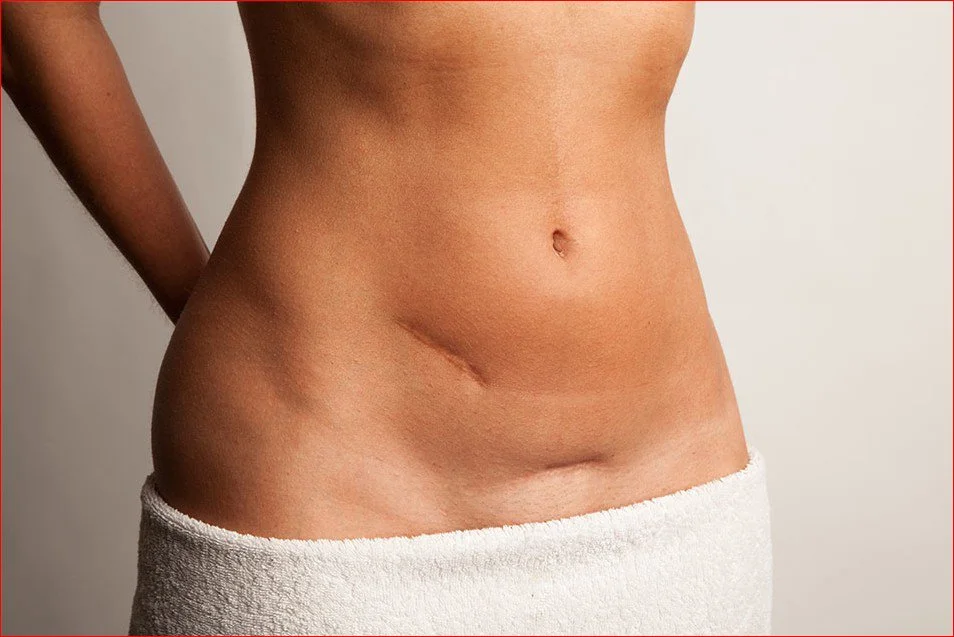
| Metric / Clinical Parameter | Small Seroma (<30 mL) | Moderate to Large Seroma (>30 mL) | Recurrent / Encapsulated Seroma |
| Primary Symptoms | Mild squishiness, localized tightness | Visible water-balloon bulge, sloshing feel | Firm, persistent lump (pseudocyst) |
| Treatment Approach | Body reabsorption & compression | Fine-needle aspiration in clinic | Re-aspiration, temporary drain, or sclerotherapy |
| Needle Aspiration Required | No | Yes (May require 1 to 3 sessions) | Yes (Combined with medical agents) |
| Compression Protocol | Standard stage 1 or 2 garment | Compression + targeted lipofoam pads | High-pressure compression + surgical binder |
| Average Resolution Time | 1 to 3 Weeks | 2 to 4 Weeks | 4 to 8 Weeks |
Frequently Asked Questions (FAQ)
What is the difference between a seroma and a hematoma after liposuction?
A seroma is a collection of pale yellow, clear, or straw-colored lymphatic fluid and plasma. A hematoma is a localized collection of blood beneath the skin, which typically forms rapidly within the first 24 to 48 hours post-op. Hematomas appear dark, purple, or deep red, cause firm or hard swelling, and are accompanied by sharper pain and intense skin bruising.
Can an untreated seroma turn into a permanent problem?
Yes. If a large seroma is left untreated for an extended period, the body may treat the fluid pocket as a chronic wound and form a fibrous capsule around it, creating a condition known as a pseudocyst or seroma cavity. Once a pseudocyst forms, the fluid can no longer be reabsorbed naturally or evacuated easily with a needle, often requiring minor surgical excision to flatten the skin contour.
Is it safe to perform manual lymphatic drainage (MLD) if I have an active seroma?
Yes, but the massage technique must be adapted by a certified therapist. MLD should gently direct fluid away from the seroma site toward functioning lymph nodes to encourage systemic clearance. However, therapists should avoid aggressive, heavy pressing directly over a large, tense seroma, as excessive force can cause skin irritation or push fluid into adjacent healthy tissues.
How do I know if my seroma has become infected?
An infected seroma (abscess) requires immediate medical attention. Signs of infection include increasing redness around the swelling, skin that feels hot to the touch, severe or worsening pain, foul-smelling or cloudy yellow/green discharge, and systemic symptoms like fever or chills. If you notice these signs, contact your surgeon right away for antibiotic treatment or drainage.

 |
|