Skip to content
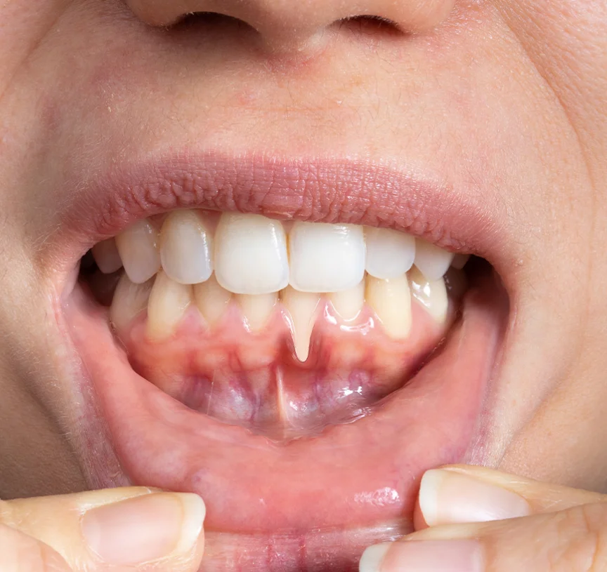
Gingival recession, the progressive loss of protective gum tissue surrounding the necks of the teeth, is an insidious oral condition affecting millions of adults worldwide. When marginal gum tissue recedes, the delicate cementum covering the tooth roots becomes directly exposed to the hostile oral environment. This loss of biological seal leads to hypersensitivity to cold and hot stimuli, root caries, aesthetic imbalance known as long tooth syndrome, and eventual tooth mobility if left unmanaged. While diligent oral hygiene and gentle brushing techniques can prevent further recession, lost keratinized gingival tissue cannot regenerate spontaneously. For definitive anatomical restoration and biological protection, a gum graft provides a reliable surgical periodontic solution.
Patients considering periodontal therapy frequently seek clinical clarification on what is a gum graft, explore the delicate microsurgical phases of gum graft surgery, review documented tissue transformations in gum graft before and after portfolios, inquire about post-operative healing milestones like gum graft one week later, and evaluate clinical variables to determine the overall gum graft cost and how much is a gum graft. This clinical guide breaks down the biological mechanisms, harvesting techniques, recovery benchmarks, and pricing dynamics.
Table of Contents
What Is a Gum Graft and Why Is It Necessary?
If you are researching what is a gum graft, it is a specialized periodontal microsurgical procedure designed to halt gingival recession, thicken thin tissue biotypes, cover exposed and sensitive root surfaces, and re-establish a resilient barrier of attached keratinized gingiva around natural teeth or dental implants.
Get Free Consultation
Have any questions? Leave your details and we'll get back to you shortly.
Gingival tissue serves as the primary mechanical and immunological barrier protecting the underlying alveolar bone:
- Mechanical Shielding: Healthy attached gingiva is tightly bound to the underlying periosteum and bone, resisting shearing forces generated during chewing and aggressive tooth brushing.
- Bacterial Defense: When recession destroys this attached band, loose alveolar mucosa abuts the tooth margin, creating deep periodontal pockets where anaerobic bacteria flourish.
- Root Sensitivity and Decay Prevention: Exposed root surfaces lack hard enamel protection; covering them restores a thermal barrier and stops acidic erosion.
- Aesthetic Harmony: Grafting restores symmetrical gum contours and resolves uneven margins across the visible smile line.
Surgical Techniques: How Gum Graft Surgery Is Performed
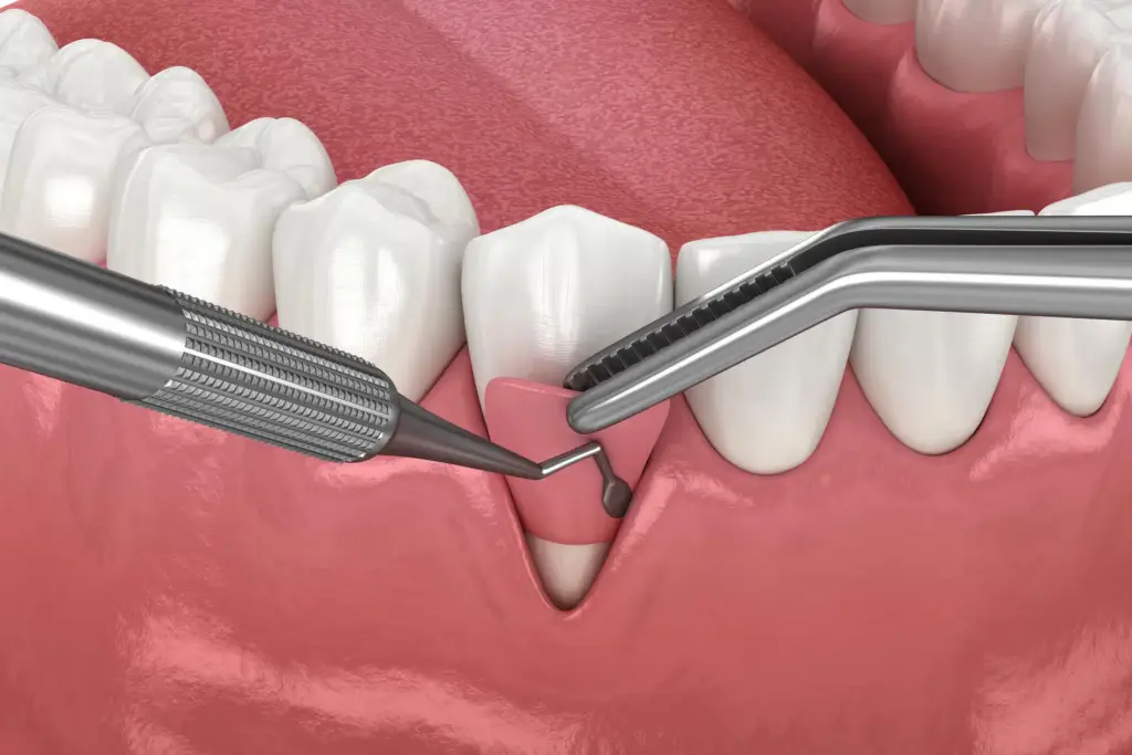
During modern gum graft surgery, periodontists utilize magnification loupes or surgical microscopes alongside micro-instruments to optimize vascular survival and ensure patient comfort. Depending on tissue thickness and the anatomy of the recession defect, surgeons select from three core grafting techniques:
1. Connective Tissue Graft (The Gold Standard)
The most common and predictable technique for covering exposed roots. The periodontist creates a subepithelial envelope or tunnel at the recipient site. A small flap is gently raised on the roof of the mouth (hard palate), and a thin layer of subepithelial connective tissue is harvested from beneath the surface mucosa. The palate flap is immediately closed, leaving the surface layer intact. The harvested graft is secured directly over the exposed tooth root and covered by the overlying gum tissue.
2. Free Gingival Graft
Used primarily to widen the band of tough, keratinized tissue rather than achieve complete root coverage. A full-thickness strip of tissue (including the surface epithelium) is harvested directly from the palate and sutured into a prepared recipient bed at the base of the receding area. This method is common for lower front teeth where muscle attachments continually pull at the gum margin.
3. Pedicle (Lateral Sliding) Graft
When thick, healthy gingival tissue remains immediately adjacent to the receded tooth, a partial flap is incised, gently shifted laterally over the exposed root, and sutured in place while maintaining its original blood supply. This technique eliminates the need for a secondary donor site on the palate.
4. Acellular Dermal Matrices (Allografts)
For patients with multiple receding teeth or those wishing to avoid a palatal donor site, bioengineered donor matrices (such as purified human donor dermis or porcine collagen matrices) are utilized. These scaffolds recruit host blood vessels and fibroblasts, transforming into native gum tissue without harvesting tissue from the palate.
Recovery Milestones: Gum Graft One Week Later
Understanding the biological healing timeline prevents complications and ensures optimal graft integration:
- Days 1 to 3 (Plasmic Circulation): The grafted tissue survives entirely through capillary diffusion from the surrounding recipient bed. Swelling and mild bruising peak around day 2 or 3. Minor oozing from the palate is managed with a protective acrylic stent or hemostatic collagen sponges. Cold packs applied externally reduce facial swelling.
- Day 4 to 6 (Revascularization): Microscopic capillary buds bridge the gap between the graft and the recipient site, establishing primary vascular perfusion.
- Gum Graft One Week Later: At gum graft one week later, early granulation tissue has anchored the graft firmly in place. The graft may appear pale pink, whitish, or sloughing on its superficial surface, which is a normal biological phenomenon representing superficial epithelial turnover. Pain at the recipient site is minimal, while the palate donor area feels like a mild pizza burn. External facial swelling has resolved, and patients can chew very soft foods comfortably on the untreated side of the mouth.
- Weeks 2 to 3: Superficial sutures are gently removed or dissolve on their own. Immature collagen fibers mature, anchoring the new tissue tightly to the tooth root.
- Months 2 to 6: Complete tissue remodeling and creeping attachment occur, creating a mature, keratinized collar of gum tissue that blends invisibly with adjacent structures.
Real-World Results: Gum Graft Before and After
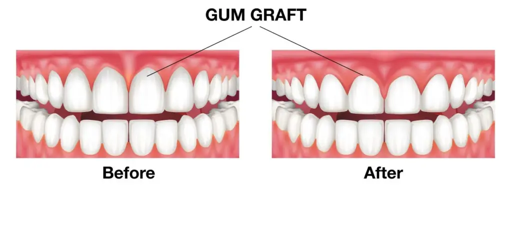
Examining documented gum graft before and after clinical portfolios illustrates substantial therapeutic and cosmetic improvements:
- Elimination of Exposed Roots: Yellowish, elongated root dentin is completely covered by thick, healthy pink tissue.
- Resolution of Tooth Sensitivity: Patients regain the ability to consume ice water, citrus fruits, and hot beverages without sharp, shooting nerve pain.
- Thickened Tissue Biotype: Thin, translucent gum tissue prone to tearing is transformed into a thick, resilient, and protective keratinized zone.
- Natural Tissue Blending: Modern tunneling techniques preserve the interdental papillae, ensuring zero visible scarring or mismatched tissue borders.
Essential Post-Operative Care and Protection
To avoid disrupting the fragile microvascular connections of the healing graft, patients must follow strict clinical guidelines:
- Strictly No Brushing or Flossing the Surgical Site: Do not touch the grafted area with a toothbrush or dental floss for at least two to three weeks. Clean the area exclusively using prescribed chlorhexidine gluconate antimicrobial mouthwash, bathing the site gently without vigorous swishing.
- Avoid Direct Mechanical Trauma: Never pull your lip or cheek outward to inspect the graft in a mirror, as tension can tear the micro-sutures and detach the graft.
- Dietary Adjustments: Maintain a soft, cool, or room-temperature diet (yogurt, scrambled eggs, smoothies, lukewarm pureed soups) for two weeks. Avoid hot liquids, hard crusts, chips, seeds, and nuts.
- No Smoking or Vaping: Nicotine causes severe peripheral vasoconstriction, starving the graft of essential oxygen and dramatically increasing the risk of complete graft necrosis.
Comparative Matrix: Gum Grafting Techniques
| Clinical Parameter | Connective Tissue Graft | Free Gingival Graft | Acellular Dermal Matrix (Allograft) |
| Primary Objective | Complete root coverage and aesthetics | Increasing keratinized tissue band | Multi-tooth coverage without palate donor |
| Palatal Donor Site | Required (Internal layer only) | Required (Surface tissue removed) | None (Bioengineered donor material) |
| Donor Site Discomfort | Mild (Palate flap is closed) | Moderate (Open surface wound heals) | Zero palatal pain |
| Root Coverage Predictability | Very High (90 to 95 percent) | Moderate | High across multiple contiguous teeth |
| Aesthetic Color Match | Seamless pink tissue blend | May heal slightly paler than adjacent gum | Excellent tissue blending |
| Healing Timeline | 2 to 4 Weeks | 3 to 5 Weeks | 3 to 4 Weeks |
What Determines the Gum Graft Cost?
When inquiring about the overall gum graft cost or asking how much is a gum graft, prospective patients will find that pricing is personalized rather than fixed. Total procedural fees depend on several specific clinical, surgical, and logistical factors:
- Number of Teeth and Recession Severity: Treating an isolated single tooth recession defect differs significantly from managing generalized recession across four to eight teeth in multiple quadrants.
- Graft Material and Harvesting Technique: Using autologous tissue harvested from the palate incurs surgeon time and specialized microsurgical instruments. Opting for advanced bioengineered matrices (allografts) introduces dedicated material and biomaterial preservation costs while eliminating palatal donor surgery.
- Surgical Complexity and Defect Classification: Miller Class 1 and Class 2 defects (where adjacent interdental bone is intact) are straightforward to correct. Miller Class 3 and 4 defects (with significant interproximal bone loss) require advanced biomaterials, such as enamel matrix derivatives (e.g., Emdogain) or bone grafts, to facilitate attachment.
- Specialist Qualifications and Surgical Tools: Board-certified periodontists with specialized residency training in periodontal plastic surgery and microsurgical tunneling platforms reflect their procedural expertise in their fee structure.
- Geographic Location and Healthcare Frameworks: Procedural costs vary markedly between domestic metropolitan private practices and dental tourism hubs like Turkey. Programs in Turkey routinely bundle periodontal workups, multi-tooth grafting, 3D imaging, prescription medications, luxury hotel stays, and private transfers into structured, cost-effective care plans.
Frequently Asked Questions (FAQ)
Can receding gums grow back naturally without surgery?
No. Once gum tissue has receded and the underlying attachment apparatus is lost, it cannot regenerate or creep back down the root on its own. While proper brushing habits and regular dental cleanings prevent the recession from worsening, surgical grafting is the only clinical method capable of physically recovering the exposed root surface and restoring lost attached gingiva.
Does gum graft surgery hurt?
During the procedure, you will feel no pain because the area is thoroughly anesthetized with local anesthesia. Conscious sedation options are also available for anxious patients. Post-operative discomfort at the graft site is generally mild and feels like a tender gum ache. If a palatal harvest was performed, the roof of the mouth will feel similar to a hot pizza burn for three to seven days, which is well-managed with prescribed pain relievers and a protective palatal stent.
What should a gum graft look like one week later?
One week after surgery, it is completely normal for the graft to look white, greyish, or patchy. This appearance often alarms patients, but it represents the normal biological sloughing of superficial epithelial cells while deeper connective tissue layers integrate. As long as there is no active bleeding, swelling, or foul odor, this is a sign of healthy revascularization.
What happens if I choose not to get a gum graft?
Leaving progressive gingival recession untreated allows the bone beneath the gum to recede alongside the soft tissue. Over time, the root becomes vulnerable to decay, sensitivity worsens, and the loss of skeletal bone support can ultimately lead to tooth looseness and premature tooth loss.

 |
|