Skip to content
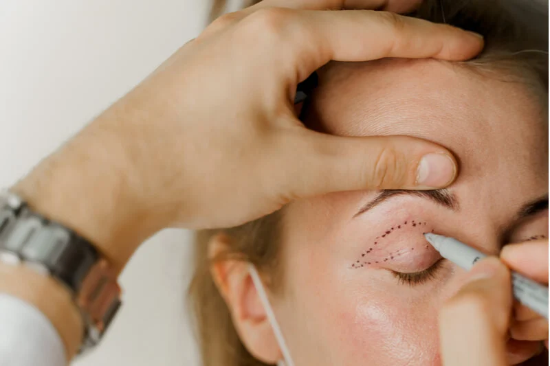
Blepharoptosis, commonly referred to as ptosis, is an anatomical condition characterized by an abnormal drooping of one or both upper eyelid margins. Unlike dermatochalasis, where excess stretched skin folds downward over the eyelashes, true ptosis stems from structural impairment, stretching, or dehiscence of the specialized muscles responsible for lifting the lid, primarily the levator palpebrae superioris. When the upper eyelid margin falls across the pupillary aperture, it obstructs the superior visual field, creates chronic ocular fatigue, forces patients into compensatory chin-up postures, and causes persistent forehead tension headaches due to chronic contraction of the frontalis muscle. While non-surgical crutch glasses offer temporary relief, ptosis surgery provides the definitive surgical cure.
Individuals researching treatment options frequently inquire about functional and cosmetic transformations in ptosis surgery before and after portfolios, evaluate the surgical steps of eyelid ptosis surgery, examine long-term scar maturation regarding a ptosis surgery scar, assess the clinical differences between muscle repair and blepharoplasty, and research the clinical variables that determine the overall ptosis surgery cost. This clinical guide examines eyelid biomechanics, surgical modalities, scar management, and pricing dynamics.
Table of Contents
What Is Eyelid Ptosis Surgery and How Does It Work?
An eyelid ptosis surgery is an ophthalmic and oculoplastic procedure designed to tighten, advance, or suspend the retracting mechanisms of the upper eyelid, elevating the resting eyelid margin back to its natural anatomical position above the pupil.
Get Free Consultation
Have any questions? Leave your details and we'll get back to you shortly.
The surgical strategy depends on evaluating two crucial pre-operative measurements:
- Marginal Reflex Distance One (MRD1): The distance measured in millimeters from the center of the pupillary light reflex to the central margin of the upper eyelid. Normal MRD1 ranges between four and five millimeters; an MRD1 below two millimeters indicates functional ptosis.
- Levator Muscle Excursion (Function): The total vertical excursion of the upper eyelid margin measured while shifting gaze from extreme downgaze to extreme upgaze, holding the brow completely still. Excellent function exceeds fifteen millimeters, while poor function measures under four millimeters.
Surgical Techniques: Matching the Procedure to Muscle Function

Oculoplastic surgeons select the surgical approach according to the patient’s levator excursion and response to pharmacological testing:
1. External Levator Aponeurosis Advancement (Transcutaneous Approach)
The gold standard technique for patients presenting with moderate to good levator muscle function (greater than eight millimeters). The surgeon makes a delicate horizontal incision within the natural upper eyelid crease. The underlying levator aponeurosis is located, freed from surrounding fibrotic attachments, advanced forward, and sutured directly onto the anterior surface of the tarsal plate. The patient is often asked to open their eyes during the operation under local anesthesia to verify intraoperative height and symmetry.
2. Posterior Conjunctival-Müller’s Muscle Resection (PMMR / Fasanella-Servat)
Indicated for mild to moderate ptosis (one to two millimeters of droop) in patients who show a positive response to topical phenylephrine eye drops during clinical evaluation. In this internal, scarless approach, the eyelid is inverted, and a precise strip of Müller’s muscle and conjunctiva is clamped and resected from the inside. Because the entire procedure is performed behind the eyelid, there is no external skin incision.
3. Frontalis Sling (Brow Suspension)
The only viable clinical solution for severe ptosis with poor to absent levator function (under four millimeters), typical in congenital ptosis or myasthenia gravis. Because the native levator muscle cannot lift the lid, the surgeon bridges the tarsal plate to the strong frontalis muscle of the forehead using biocompatible synthetic materials or an autologous fascia lata graft taken from the thigh. The patient then uses their eyebrow muscles to elevate the eyelid.
Incision Management: Healing of a Ptosis Surgery Scar
Understanding the lifecycle of a ptosis surgery scar helps patients maintain realistic post-operative expectations:
- Days One to Seven: For external approaches, the incision sits directly in the supratarsal crease. Skin edges are closed with microscopic sutures, which are removed five to seven days after surgery. The scar line will appear pink and mildly elevated.
- Weeks Two to Six: As inflammatory collagen undergoes early cross-linking, the scar may feel slightly firm to the touch. Because eyelid skin is among the thinnest tissues on the human body with exceptional vascularity, hypertrophic or keloid scarring is extremely rare.
- Months Three to Twelve: The incision line matures into a faint, pale line that settles into the natural eyelid fold. When the eyes are open, the scar is hidden within the crease; when the eyes are closed, it appears as a subtle, natural skin line.
- For Internal Approaches: Patients undergoing posterior Müller’s muscle resections have zero external cutaneous scars.
Functional and Aesthetic Outcomes: Ptosis Surgery Before and After
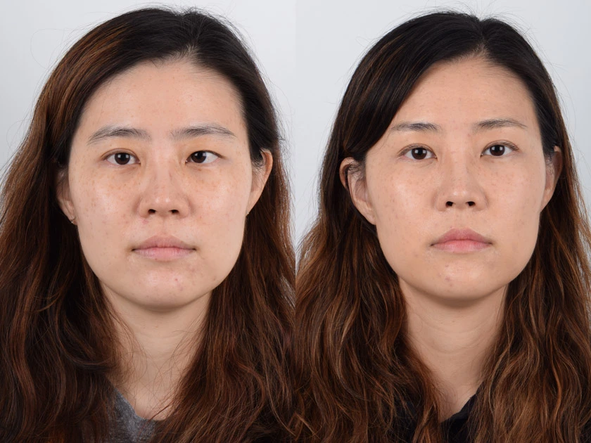
Examining clinical ptosis surgery before and after portfolios demonstrates significant improvements across daily function and aesthetics:
- Restoration of Full Peripheral Vision: Eliminating the heavy lid curtain opens the superior visual field, allowing patients to drive safely, read without strain, and look upward effortlessly.
- Disappearance of Forehead Fatigue: The constant urge to raise the eyebrows to see disappears, resolving forehead muscle strain and smoothing out chronic horizontal forehead wrinkles.
- Symmetrical, Awake Appearance: Drooping, asymmetrical, sleepy-looking eyes are realigned into balanced, alert contours with natural pupillary exposure.
- Combined Eyelid Tightening: When paired with upper blepharoplasty, redundant hooded skin is removed concurrently with muscle advancement, creating a smooth upper lid contour.
Essential Post-Operative Recovery Guidelines
- Cold Compresses Diligently: Apply chilled gel packs or clean ice wrapped in sterile gauze over the closed eyelids for ten to fifteen minutes every waking hour during the first forty-eight hours to control swelling.
- Lubricating Ointments and Drops: Temporary lagophthalmos (an inability to close the eye completely) can occur during the first week due to localized muscle swelling. Use prescribed artificial tears during the day and thick lubricating eye ointment at night to prevent corneal exposure.
- Strictly Avoid Bending and Lifting: Refrain from heavy lifting (over ten pounds), vigorous exercise, and bending down so your head is below your heart for at least two weeks to prevent hematoma formation.
- Keep the Incision Clean and Dry: Avoid soaking the surgical site in water or rubbing the eyes for two weeks. Clean the lid margin with sterile saline and apply antibiotic ointment as directed.
Comparative Matrix: Ptosis Surgery vs. Upper Blepharoplasty
| Clinical Parameter | External Levator Repair | Internal Müller’s Resection | Upper Blepharoplasty Alone |
| Primary Pathology | Weak or stretched levator muscle | Mild muscle weakness / Horner’s | Excess skin and herniated fat |
| Target Tissue | Muscle and aponeurosis | Müller’s muscle and conjunctiva | Redundant skin and orbicularis muscle |
| Effect on Lid Height | Directly raises the eyelid margin | Raises margin by 1.5 to 2 mm | Does not raise the lid margin |
| Incision Location | Hidden in eyelid crease | Inside eyelid (no skin cut) | Hidden in eyelid crease |
| External Scar | Yes (hidden in natural crease) | Zero external scar | Yes (hidden in natural crease) |
| Visual Field Benefit | Clears pupillary axis directly | Clears pupillary axis | Removes peripheral skin weight |
| Typical Downtime | 10 to 14 Days | 5 to 7 Days | 7 to 10 Days |
What Determines the Ptosis Surgery Cost?
When researching the ptosis surgery cost, prospective patients will discover that total fees are personalized rather than standardized. Several anatomical and surgical factors influence pricing:
- Unilateral vs. Bilateral Correction: Treating a single drooping eye requires delicate adjustments to match the contralateral lid, while bilateral cases require double the operating room resources.
- Surgical Technique Complexity: An internal conjunctival resection requires less operative time than an extensive external levator advancement or a complex frontalis sling procedure.
- Need for Concurrent Blepharoplasty: Ptosis repair is frequently combined with an upper blepharoplasty to remove overlapping dermatochalasis, which adds surgical steps.
- Anesthesia Preference: Procedures performed under local anesthesia with light oral sedation involve lower facility fees than cases requiring deep intravenous sedation or general anesthesia.
- Surgeon Credentials and Oculoplastic Specialization: Board-certified oculoplastic surgeons (ophthalmologists with subspecialty fellowship training in eyelid and orbital plastic surgery) reflect their precision and lower revision rates in their fee structures.
- Geographic Location and Treatment Packages: Healthcare overhead creates notable price variations between private metropolitan clinics in the UK or US and accredited healthcare destinations like Turkey. Programs for ptosis surgery in Turkey frequently bundle ophthalmological diagnostics, the surgery, protective drops, boutique hotel stays, and private VIP transfers into structured care packages.
Frequently Asked Questions (FAQ)
What is the difference between ptosis surgery and an upper blepharoplasty?
An upper blepharoplasty removes only redundant, sagging skin and excess fat from the eyelid; it does not change the position of the actual eyelid margin. If a patient with a weak levator muscle undergoes only a blepharoplasty, the eyelid margin will remain droopy. Ptosis surgery specifically shortens or advances the underlying muscle to physically raise the eyelid margin above the pupil.
Will I be awake during ptosis surgery?
Yes, external levator advancement is most commonly performed under local anesthesia with light sedation. Because adjusting the eyelid height demands millimeter-level accuracy, the surgeon frequently asks you to open and close your eyes during the procedure to evaluate contour and symmetry against the opposite eye.
Can ptosis surgery cause dry eye symptoms?
Mild, temporary dry eye symptoms are common during the first two to three weeks because the newly elevated eyelid exposes more of the ocular surface to air, and local tissue swelling can temporarily delay complete blink closure. Prescribed lubricating drops during the day and ophthalmic ointments at bedtime prevent corneal dryness until natural blinking normalizes.
Is ptosis surgery permanent?
Yes, surgical reattachment or advancement of the levator muscle provides long-lasting structural correction for decades. While the eyelid tissues will continue to experience natural cellular aging over time, the surgical tightening does not simply wear off, and recurrent droop requiring revision is rare when performed by experienced oculoplastic specialists.

 |
|