Skip to content

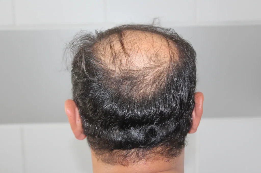
Modern follicular restoration techniques such as Follicular Unit Extraction (FUE) and Direct Hair Implantation (DHI) have transformed surgical hair restoration from invasive strip incisions into micro-punch precision. Because the occipital and parietal areas at the back and sides of the head are genetically programmed with 5-alpha reductase resistance, these follicles serve as the primary reservoir for grafting balding zones. However, the donor supply is finite. When surgical planning fails to calculate density ratios or uncertified technicians prioritize unrealistic graft counts, patients often face visible donor area thinning.
Patients experiencing an uneven, depleted back of the head frequently seek clinical clarity on hair transplant donor area thinning, research the distinction between temporary shedding and permanent loss regarding donor area thinning after hair transplant, evaluate the severity of an overharvested donor area hair transplant, look for actionable solutions on how to fix donor area thinning, differentiate between donor area shock loss vs overharvesting, and map their clinical timeline during donor area thinning recovery. This comprehensive clinical guide examines safe extraction capacities, physiological mechanisms, surgical repair modalities, and cost determinants.
Table of Contents
What Causes Donor Area Thinning After Hair Transplant?
Understanding the primary causes of follicular depletion in the harvesting zone involves differentiating between temporary biological trauma and permanent structural depletion:
Get Free Consultation
Have any questions? Leave your details and we'll get back to you shortly.
- Surgical Overharvesting (Depletion of Follicular Density): In an uncompromised occipital scalp, native density ranges between sixty-five and eighty-five follicular units per square centimeter. A safe extraction protocol dictates taking no more than fifteen to twenty percent of the available donor pool in a single session (roughly one out of every four to five follicular units). When aggressive clinics harvest forty to fifty percent of adjacent follicles to hit excessive mega-session promises, the remaining spacing collapses, exposing the scalp.
- Donor Area Shock Loss (Telogen Effluvium): During micro-punch extraction, localized tissue vibration, mechanical micro-trauma, tumescent fluid infiltration, and disruption of capillary perfusion trigger a defensive response. Surrounding unharvested follicles enter a premature resting (telogen) phase. This produces a temporary, diffuse patchiness that mimics permanent overharvesting between weeks two and eight post-surgery.
- Inappropriate Micro-Punch Caliber: Using extraction punches wider than zero point nine millimeters leaves larger circular extraction wounds. As these sites heal with microscopic fibrotic scar tissue, the contracture pulls surrounding hair-bearing skin taut, reducing natural hair grouping coverage.
- Clustered and Non-Homogeneous Extraction Patterns: Safe harvesting requires skipping adjacent follicular units across a wide surface area. If a technician repeatedly harvests adjacent follicular units from a narrow focal zone, it creates localized, completely barren circular holes known as a moth-eaten appearance.
- Subclinical Scalp Conditions: Unaddressed pre-existing conditions, including retrograde androgenetic alopecia (thinning that creeps upward from the nape of the neck) or diffuse unpatterned alopecia (DUPA), mean the donor zone itself is sensitive to dihydrotestosterone (DHT) and prone to thinning over time.
Differentiating the Causes: Shock Loss vs. Overharvesting
Determining whether thinness at the back of the head is temporary or permanent is essential for establishing an appropriate treatment plan:
- Onset and Progression: Shock loss develops rapidly between two to six weeks after surgery, presenting as a soft, diffuse thinning without distinct edges. True overharvesting is present immediately once post-operative scabs shed around day ten and remains stationary over months.
- Presence of Follicular Openings: Under high-magnification trichoscopy, areas affected by shock loss retain visible, empty follicular pores containing dormant hair roots beneath the skin. Overharvested areas exhibit total absence of follicular apertures, replaced by pinpoint hypopigmented circular scars.
- Scalp Texture and Fibrosis: Scalps with shock loss remain pliable, soft, and elastic. Severely overharvested zones feel firm, fibrotic, bound-down, and irregular due to dense subcutaneous scarring from repeated punch passes.
Clinical Protocols: How to Fix Donor Area Thinning
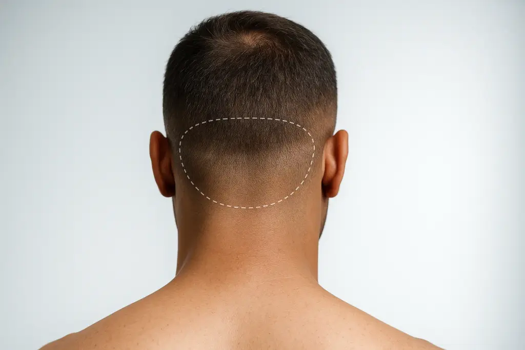
Repairing a depleted, patchy donor area requires specialized reconstructive hair restoration expertise. Clinicians utilize several corrective options:
1. Scalp Micropigmentation (SMP)
The most reliable, non-surgical camouflage modality for overharvested donor areas:
- Medical Pigment Implantation: A specialized medical practitioner uses microscopic needles to deposit medical-grade, carbon-based pigments into the upper papillary dermis.
- Visual Illusion: By replicating the visual appearance of thousands of natural, shaven single-hair follicular dots, SMP eliminates the contrast between white fibrotic scars and bare scalp skin, restoring the optical appearance of uniform density under short haircuts.
2. Beard-to-Scalp FUE Transplantation
When actual hair volume must be physically added back to a depleted occipital scalp:
- Submental Follicular Harvesting: Coarse, robust single-hair follicular units are extracted from beneath the jawline and neck (beard shadow). Because beard hair has a thicker shaft diameter, fewer grafts are needed to produce optical coverage.
- Strategic Implantation: These beard grafts are implanted into the completely bare punch scars at the back of the head, breaking up linear patchiness and providing tactile coverage.
3. Autologous Regenerative Therapies (PRP, PRF, and Exosomes)
Indicated primarily for cases involving shock loss or compromised microcirculation:
- Platelet-Rich Fibrin (PRF) and Growth Factors: Injecting concentrated autologous growth factors directly into the thinned donor beds stimulates dormant shock loss follicles, promotes angiogenesis (new capillary development), and accelerates the return to the anagen growth phase.
4. Conservative Medical Stabilization
Administering oral or topical minoxidil alongside low-dose finasteride improves capillary perfusion, shortens the telogen phase of shocked follicles, and prevents native donor hair miniaturization if retrograde thinning is suspected.
Recovery Roadmap: Donor Area Thinning Recovery Timeline
Monitoring tissue changes over time helps patients assess healing progression:
- Days One to Ten (Post-Surgical Healing): The donor zone is covered with tiny, pinhead-sized crusts. Mild tightness, burning sensations, and oozing are common. Crusts naturally wash away during specialized gentle washes between days seven and ten.
- Weeks Two to Eight (The Peak Shock Loss Window): Hair shafts in and around the donor site shed. The back of the head may look patchy, uneven, or thin. This is the stage where anxiety peaks; distinguishing shock loss from overharvesting is crucial.
- Months Three to Four (Early Awakening): Dormant shocked follicles reactivate. Fine, wispy hairs begin emerging from previously bare patches, softening the transition zones.
- Months Six to Nine (Density Restabilization): Hair shafts gain normal caliber, length, and natural pigment. If thinning persists without improvement at month nine, the remaining deficit reflects permanent overharvesting that requires SMP or beard graft repair.
- Month Twelve (Final Donor Evaluation): Tissue remodeling is complete. The definitive donor density is documented, and reconstructive measures can be safely initiated.
Before and After: Reconstructive Results
Reviewing documented repair cases illustrates realistic improvements:
- Elimination of the Moth-Eaten Pattern: Patchy, see-through zones are filled or camouflaged, replacing isolated voids with balanced coverage.
- Concealment of White Dot Scars: Hypopigmented micro-scars are blended using medical-grade SMP so they do not show through surrounding hair.
- Freedom in Grooming: Patients can trim their hair to short fade lengths (grade two or three) without exposing distinct surgical punch patterns.
- Restored Scalp Health: Relieving tight, fibrotic bands through regenerative therapies softens the scalp, easing chronic tension and hypersensitivity.
Essential Donor Preservation Guidelines
- Respect the Mathematical Safe Donor Limit: Never exceed an extraction ratio of twenty percent of your native donor hair in a single surgery. A safe first session rarely exceeds three thousand to four thousand grafts, regardless of how large the balding area is.
- Verify Punch Sizes and Surgeon Involvement: Ensure your surgical team uses punches between zero point seven and zero point eight five millimeters. Verify that a licensed doctor performs or directly supervises the extraction step.
- Avoid Scratching the Healing Donor Site: In the first two weeks, donor itching can be intense due to histamine release. Scratching can tear healing micro-punctures and introduce bacteria. Apply prescribed soothing saline sprays or aloe vera gels instead.
- Shield from Direct Sun: Keep the recovering back of the head protected from direct ultraviolet radiation for at least six months by wearing a loose, breathable cap to prevent post-inflammatory hyperpigmentation.
Comparative Matrix: Donor Area Restoration Modalities
| Clinical Parameter | Scalp Micropigmentation (SMP) | Beard Hair-to-Scalp Grafting | Autologous PRF / PRP | Topical / Oral Vasodilators |
| Primary Indication | Severe overharvesting & visible scars | Deep bare gaps requiring actual hair | Shock loss & slow circulation | Diffuse shocked native hairs |
| Treatment Modality | Medical pigment tattoo | Micro-FUE surgical grafting | Autologous blood injections | Daily prescription medication |
| Number of Sessions | 2 to 3 Sessions | 1 Reconstructive surgical session | 3 to 4 Monthly sessions | Continuous daily routine |
| Discomfort Level | Mild to moderate | Mild (under local anesthesia) | Mild (micro-needling feel) | None |
| Downtime | 2 to 3 Days (mild redness) | 7 to 10 Days (scab healing) | Zero downtime | Zero downtime |
| Permanence | 3 to 5 Years (touch-ups needed) | Permanent (beard follicles) | Requires 6-12 month boosters | Active only while used |
| Risk of Complications | Pigment fading or blurring | Folliculitis, donor scarring under chin | Mild temporary tenderness | Scalp dryness, initial shedding |
What Determines the Cost of Repairing Donor Area Thinning?
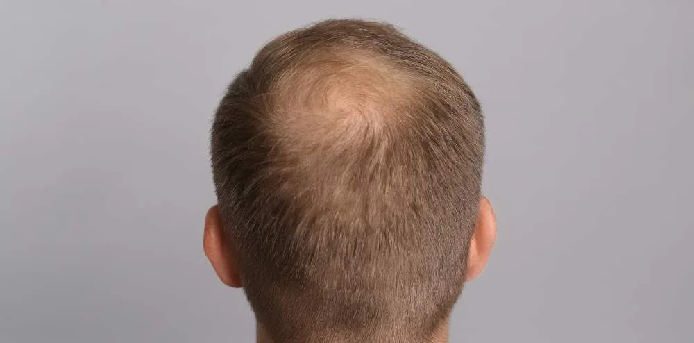
The total investment required to correct a depleted donor area varies based on individual clinical variables rather than a single fixed rate:
- Selection of Corrective Modality: A non-invasive course of scalp micropigmentation involves a fundamentally different fee structure than a complex reconstructive micro-FUE procedure utilizing delicate beard extractions.
- Extent of Surface Depletion: Treating a small, isolated moth-eaten patch at one temple corner requires fewer resources than reconstructing the entire occipital and parietal donor zone from ear to ear.
- Need for Staged Interventions: Severe cases frequently demand a multi-step protocol: first using regenerative PRP or polynucleotide therapies to re-vascularize scar tissue, followed by beard hair grafting, and finishing with SMP dotting.
- Clinical Practitioner Specialization: Board-certified hair restoration surgeons who specialize in reconstructive revisions and master SMP technicians charge fees reflective of their corrective expertise and clinical safety records.
- Geographic Location and Package Offerings: Operating overhead and clinical costs create marked price differences between private revision clinics in North America or Western Europe and internationally accredited centers in Turkey. Reconstructive packages in Turkey frequently bundle advanced digital trichoscopy, restorative beard-to-scalp grafting, hyperbaric oxygen therapy, hotel accommodation, and private VIP transfers into structured care arrangements.
Frequently Asked Questions (FAQ)
Can hair grow back in the exact punch holes where donor follicles were taken?
No. Once a follicular unit is physically extracted from the scalp with its dermal papilla and surrounding tissue, that specific follicle is permanently gone and cannot regenerate. The goal of safe surgery is to harvest only a percentage of hairs so that neighboring undisturbed follicles grow out and cover the microscopic extraction gap.
How can I tell if my thin donor area is due to temporary shock loss or permanent overharvesting?
The key indicators are timing and trichoscopic appearance. Shock loss typically appears between weeks two and six, showing empty pores where hair roots are sleeping, and the scalp remains soft and elastic. Overharvesting is visible immediately once scabs clear around day ten, characterized by missing pores, white scar tissue, and an uneven pattern that does not improve after six months.
Can beard hair be safely used to fill in a thin donor area?
Yes. Beard hair harvested from under the chin and neck serves as an excellent donor source for repairing overharvested scalps. Because beard hair shafts are naturally thicker than scalp hairs, they provide exceptional optical density when placed into bald punch scars at the back of the head, preserving remaining scalp hair.
When is it safe to undergo a repair procedure after noticing donor area thinning?
You should wait at least eight to twelve months following your initial surgery before undergoing any surgical revision or SMP. This waiting window allows all temporary shock loss to resolve completely, gives local micro-scars time to mature and soften, and lets your specialist clearly identify permanent deficits.

 |