Skip to content

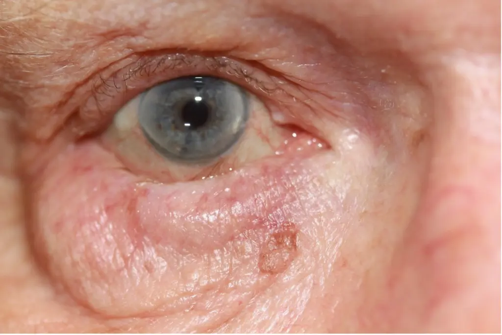
The eyelids serve as the primary protective shield for the human eye, distributing the tear film evenly with every blink while sweeping foreign particulates off the ocular surface. When the delicate structural balance of the eyelid margin fails, the lid can roll inward toward the globe, a clinical condition diagnosed as entropion. As the eyelid margin inverts, the eyelashes (cilia) and outer skin rub continuously against the ultra-sensitive cornea and bulbar conjunctiva. What begins as a minor foreign-body sensation can rapidly escalate into severe corneal abrasions, microbial keratitis, permanent corneal scarring, and preventable vision loss.
Patients experiencing involuntary eyelid inversion urgently investigate what is entropion, identify hallmark entropion sypmtoms, explore biological and anatomical entropion causes, evaluate clinical differences in entropion vs ectropion, assess non-surgical options for entropion treatment, prepare for definitive correction via entropion surgery, seek specialist care for an irritated entropion eye, and research clinical variables that dictate the overall entropion surgery cost. This clinical guide examines eyelid biomechanics, surgical correction protocols, recovery milestones, and treatment pricing dynamics.
Table of Contents
What Is Entropion and How Does It Develop?
If you are researching what is entropion, it is clinically defined as an inversion of the eyelid margin where the lash line folds inward toward the eyeball. While it can theoretically affect either lid, it most commonly involves the lower eyelid due to gravitational and anatomical predispositions.
Get Free Consultation
Have any questions? Leave your details and we'll get back to you shortly.
The mechanical failure of the eyelid margin involves four primary anatomical defects:
- Horizontal Eyelid Laxity: With advancing age, the medial and lateral canthal tendons stretch and lose their tensile firmness. The fibrous tarsal plate, the dense structural backbone of the eyelid, becomes loose, allowing the eyelid to buckle under muscular tension.
- Attenuation or Disinsertion of Lower Eyelid Retractors: The capsulopalpebral fascia and inferior tarsal muscle act as the downward anchors of the lower lid. When these retractor bands stretch, tear, or detach from the inferior tarsal border, the lower edge of the tarsal plate loses its downward stability, allowing the upper lid margin to pivot inward.
- Overriding of the Preseptal Orbicularis Oculi Muscle: During blinking or involuntary squeezing, the preseptal segment of the circular orbicularis muscle rides upward over the pretarsal segment, physically rolling the lid margin toward the cornea.
- Enophthalmos and Loss of Orbital Volume: Age-related atrophy of retrobulbar fat cushions causes the eyeball to sink slightly backward into the bony orbit. This structural loss removes the normal structural support that keeps the eyelid flush, leaving space for the eyelid to rotate inward.
Structural and Biological Triggers: Entropion Causes
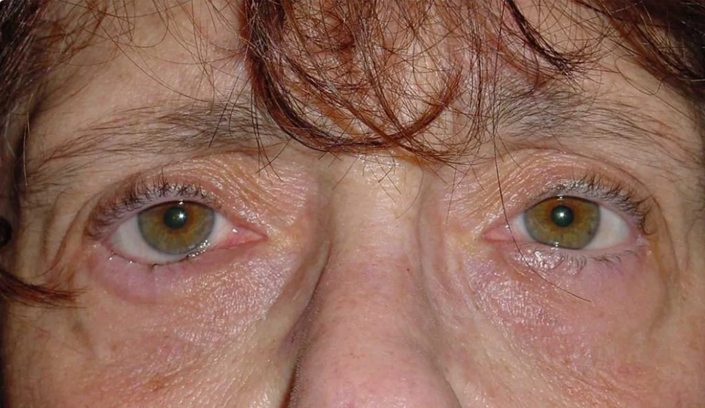
Understanding entropion causes requires categorizing the condition into distinct clinical subtypes:
- Involutional Entropion: The most prevalent form, caused by age-related tissue degradation, loss of elastic fibers, canthal tendon stretching, and retractor disinsertion in older adults.
- Cicatricial Entropion: Caused by structural scarring and contracture of the inner palpebral conjunctiva. Common triggers include ocular cicatricial pemphigoid, Stevens-Johnson syndrome, chemical burns, trachoma infections, and previous eyelid trauma.
- Spastic Entropion: Triggered by intense ocular surface inflammation, corneal injury, or recent ocular surgery. The persistent pain induces severe, sustained spasms of the orbicularis oculi muscle, forcing the eyelid inward.
- Congenital Entropion: A rare developmental anomaly present at birth, resulting from tarsal plate hypoplasia, congenital retractor defects, or hypertrophic pretarsal muscle bands, distinct from benign epiblepharon.
Identifying the Clinical Signs: Entropion Symptoms
Living with an inverted entropion eye produces progressive discomfort. The hallmark entropion sypmtomps include:
- Persistent Foreign Body Sensation: Patients report feeling as though an eyelash, grain of sand, or piece of glass is trapped in the eye with every blink.
- Chronic Epiphora (Excessive Tearing): Mechanical friction from eyelashes stimulates the lacrimal gland via trigeminal reflex loops, producing non-stop watery tears.
- Severe Photophobia and Blepharospasm: Inward-turning lashes irritate corneal nerve endings, making bright indoor and outdoor light intolerable and inducing involuntary squinting.
- Conjunctival Hyperemia and Mucous Discharge: Continuous mechanical irritation causes the white sclera to appear bloodshot, accompanied by thick, sticky mucous discharge.
- Corneal Micro-Scratches and Neovascularization: Uncorrected friction produces punctate epithelial erosions. If neglected, these scratches advance into non-healing corneal ulcers, bacterial keratitis, and vascular pannus growth that clouds central vision.
Differential Diagnosis: Entropion vs Ectropion
Patients frequently confuse these two opposing eyelid malpositions. Comparing entropion vs ectropion clarifies their fundamental differences:
| Clinical Parameter | Entropion | Ectropion |
| Direction of Margin | Margin rolls inward toward the eye | Margin sags and turns outward away from the eye |
| Lash Contact | Eyelashes rub directly against cornea | Eyelashes point away; conjunctiva is exposed |
| Primary Pathology | Canthal laxity, retractor disinsertion, muscle override | Canthal laxity, orbicularis denervation, facial palsy |
| Primary Risk | Corneal scratching, ulcers, and blindness | Severe ocular desiccation, epiphora, conjunctival keratinization |
| Cosmetic Appearance | Eyelashes appear hidden or missing | Red, raw inner eyelid lining is visibly exposed |
| Surgical Approach | Retractor reattachment & muscle repositioning | Horizontal lid shortening & tarsal strip suspension |
Non-Surgical and Temporary Measures: Entropion Treatment
While definitive correction requires realignment, conservative entropion treatment stabilizes the ocular surface while awaiting surgery:
1. Medical Lubrication and Barrier Ointments
Applying preservative-free artificial tears by day and high-viscosity petroleum-based ophthalmic ointments at bedtime coats the cornea in a protective lubricating film, reducing mechanical friction from inverted eyelashes.
2. Therapeutic Soft Contact Lenses (Bandage Lenses)
A large-diameter, plano bandage contact lens acts as a physical shield over the corneal epithelium. The inverted lashes rub harmlessly against the silicone hydrogel lens rather than the living cornea, relieving pain instantly.
3. Eyelid Taping (Quickert Taping)
A small strip of hypoallergenic medical tape is placed near the lower lash line and pulled downward onto the upper cheek. This physical tension keeps the eyelid margin rolled outward temporarily.
4. Botulinum Toxin Injections
For spastic entropion, micro-doses of botulinum toxin injected into the pretarsal and preseptal orbicularis muscle temporarily paralyze the hyperactive fibers. This stops muscular override for three to four months, giving corneal abrasions time to heal.
Definitive Realignment: Entropion Surgery
When anatomical tissue defects are present, entropion surgery represents the gold standard curative intervention, completed as an outpatient procedure under local anesthesia with light oral sedation:
1. Lateral Tarsal Strip Procedure (Horizontal Tightening)
The surgeon makes a small lateral canthotomy incision at the outer corner of the eye, exposes the lateral border of the tarsal plate, shortens it by a measured distance, and anchors it directly to the inner periosteum of the lateral orbital rim (Whitnall’s tubercle). This resolves horizontal eyelid laxity permanently.
2. Lower Eyelid Retractor Reattachment (Jones / Quickert / Wies Procedure)
Through a subciliary incision placed just beneath the lower lash line, the surgeon identifies the detached capsulopalpebral fascia and re-anchors it firmly to the inferior border of the tarsal plate with fine, non-absorbable sutures. This restores the downward stabilizing pull, preventing the margin from rotating upward.
3. Orbicularis Muscle Resection
A narrow horizontal strip of the preseptal orbicularis muscle is excised to prevent it from sliding upward over the tarsus during blinking.
4. Transverse Tarsotomy and Marginal Eversion (Wies Procedure)
Indicated for cicatricial entropion. The surgeon incises through the full thickness of the eyelid four millimeters below the margin, placing rotational eversion sutures that mechanically tilt the scarred eyelid margin outward. In severe cases, a mucous membrane graft (harvested from the inside of the lip or hard palate) is placed to replace contracted conjunctival tissue.
Recovery Roadmap: What to Expect After Eyelid Surgery
Post-operative recovery is straightforward and predictable:
- Days One to Three (Acute Swelling and Bruising): An eye pad may be worn for the first twenty-four hours. Mild bruising around the lower lid and cheek peaks on day two or three. Cold gel packs applied for ten minutes every waking hour soothe swelling. Patients instill prescribed antibiotic eye ointments twice daily along the suture line.
- Days Four to Seven (Suture Removal): External non-absorbable skin sutures are removed at the clinic between day five and seven. Swelling subsides, and the foreign-body sensation is gone because the lashes no longer touch the cornea.
- Weeks Two to Four (Return to Full Activity): Residual discoloration fades completely. Patients resume light fitness routines, wear makeup, and return to standard office jobs within ten to fourteen days.
- Months One to Three (Final Tissue Stabilization): Deep anatomical healing completes, leaving a discreet, faint scar hidden beneath the lower lash line. Eyelid excursion and tear distribution normalize completely.
Visual Transformations: Before and After Realignment
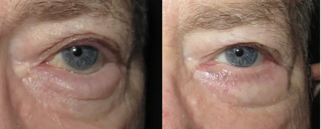
Reviewing documented clinical cases illustrates critical functional and aesthetic improvements:
- Immediate Corneal Relief: The mechanical scraping of eyelashes against the cornea ceases the moment the patient leaves the operating theater.
- Re-Established Lash Architecture: Eyelashes emerge outward in a natural, forward-facing orientation.
- Resolution of Chronic Redness: Constant ocular tearing, bloodshot sclera, and morning crusting clear within days of surgery.
- Preservation of Visual Acuity: Eliminating persistent corneal friction halts ulceration, preventing permanent corneal opacities and vision loss.
Essential Post-Operative Safety Guidelines
- Apply Antibiotic Ophthalmic Ointment Religiously: Use your prescribed antibiotic ointment inside the lower eye fornix and along the surgical incision line as directed to prevent infection and keep the wound hydrated.
- Absolutely No Eye Rubbing: Rubbing the healing eye during the first month can snap delicate internal anchoring sutures, causing the lid to revert to its inverted position.
- Sleep with the Head Elevated: Use two firm pillows to keep your head above heart level for the first five nights to speed lymphatic drainage and minimize morning eyelid edema.
- Protect Eyes from Dust and UV Light: Wear wrap-around sunglasses outdoors to shield the healing eyelid tissues from wind-borne debris, particulate irritation, and ultraviolet pigmentation.
Comparative Matrix: Entropion Treatment Modalities
| Clinical Parameter | Lubricants & Bandage Lenses | Botulinum Toxin Injections | Lateral Tarsal Strip & Retractor Repair | Full-Thickness Transverse Tarsotomy |
| Primary Indication | Mild symptoms & temporary relief | Spastic entropion & poor surgical candidates | Standard involutional (age-related) entropion | Severe cicatricial (scarring) entropion |
| Primary Goal | Protects cornea from lash friction | Paralyzes overriding orbicularis muscle | Corrects horizontal laxity & retractor detachment | Mechanically levers scarred lid outward |
| Invasiveness | Completely non-invasive | Minimally invasive (needle injections) | Outpatient surgical reconstruction | Outpatient full-thickness surgical cut |
| Procedural Duration | Daily home application | 5 to 10 Minutes | 30 to 45 Minutes per eyelid | 45 to 60 Minutes per eyelid |
| Discomfort Level | None | Mild pinch | Very mild (under local anesthesia) | Very mild (under local anesthesia) |
| Downtime | Zero downtime | Zero downtime | 7 to 10 Days (bruising and swelling) | 10 to 14 Days (bruising and swelling) |
| Longevity of Results | Only while in place | 3 to 4 Months (temporary) | Permanent (over 90 to 95 percent success) | Permanent structural correction |
What Determines the Entropion Surgery Cost?
When evaluating the financial investment for entropion surgery, prospective patients will discover that surgical pricing varies according to personalized anatomical and logistical variables rather than a single fixed fee:
- Unilateral vs. Bilateral Surgery: Repairing an inverted lid on a single eye involves different operating theater time and consumable surgical packs than performing simultaneous bilateral lid realignments.
- Underlying Etiology and Surgical Complexity: An involutional case treated with a standard lateral tarsal strip and retractor tightening requires less operative time than a complex cicatricial case requiring scar contracture release and mucous membrane grafting harvested from the mouth.
- Concomitant Ophthalmic Procedures: Entropion surgery is frequently paired with blepharoplasty (to remove redundant skin), repair of co-existing ectropion on the opposing lid, or tear duct probing to resolve secondary chronic tearing, adjusting operating theater fees.
- Surgeon Credentials and Specialization: Board-certified oculoplastic surgeons (ophthalmologists with dedicated multi-year subspecialty fellowship training in cosmetic and reconstructive plastic surgery of the eye) command professional fees reflecting their microsurgical precision and low recurrence rates.
- Anesthesia and Facility Standards: Procedures performed in a dedicated surgical suite with monitored conscious intravenous sedation involve different clinical overhead than minor repairs completed under straight local infiltration in a clinic room.
- Geographic Location and Healthcare Travel Packages: Operating facility overhead, liability frameworks, and currency exchange rates create marked price differences between private practices in Western Europe or North America and internationally accredited medical travel centers in Turkey. Ophthalmic surgical packages in Turkey frequently bundle pre-operative slit-lamp evaluations, high-resolution corneal imaging, the surgical procedure, all post-operative antibiotic and lubricating eye medications, boutique hotel lodging for five to seven nights, and private VIP transfers into structured care arrangements.
Frequently Asked Questions (FAQ)
Can entropion heal on its own without surgery?
No. Involutional, cicatricial, and congenital entropion are caused by permanent mechanical failures, such as stretched tendons, torn retractor muscles, and contracted scar tissue. These anatomical defects cannot self-correct. While spastic entropion may temporarily resolve once an underlying infection or inflammation clears, structural entropion requires surgical realignment to prevent permanent corneal blindness.
What happens if an entropion eye is left untreated?
Leaving an inverted eyelid untreated leads to severe complications. The constant abrasive scraping of eyelashes against the cornea strips away the protective epithelial barrier, leading to recurrent corneal abrasions, bacterial corneal ulcers, deep stromal scarring, and permanent reduction in visual acuity. In severe cases, unchecked corneal infections can cause globe perforation.
Is entropion surgery painful?
The procedure is virtually painless. The surgeon administers local anesthesia into the eyelid, numbing the operative zone completely within seconds. During the surgery, you may feel light pressure or gentle touch, but zero sharp pain. Following the procedure, mild post-operative soreness resembles a light bruise and is readily managed with standard over-the-counter paracetamol for forty-eight hours.
How quickly does vision recover after entropion surgery?
Functional visual comfort improves almost immediately because the inverted lashes no longer abrade the cornea. However, mild blurriness is common for the first five to seven days due to post-operative eyelid swelling and the temporary use of thick antibiotic eye ointments. Crisp vision stabilizes once sutures are removed and the cornea fully re-epithelializes within two to three weeks.

 |