
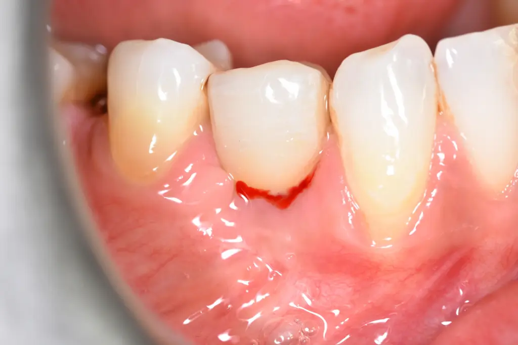
Dental implants are recognized as the gold standard for replacing missing teeth, boasting long-term success rates exceeding 95 percent. However, like natural teeth, implants rely on healthy supporting tissues to maintain stability. When harmful bacteria accumulate around an implant, they trigger an inflammatory condition known as peri-implant disease.
Understanding what is peri-implantitis is crucial for preserving your dental restorations. Patients frequently seek information about recognizing peri-implantitis symptoms, managing early peri-implantitis, evaluating the clinical peri-implantitis stages, identifying lifestyle peri-implantitis risk factors, and exploring advanced peri-implantitis treatment pathways such as laser treatment of peri-implantitis Turkey. This clinical guide provides an in-depth breakdown of how peri-implantitis develops, how to detect early warning signs, and how modern regenerative and laser therapies save compromised implants.
Table of Contents
What Is Peri-Implantitis?
To answer what is peri-implantitis, we must look at how bacterial biofilms interact with the titanium or zirconia surface of a dental implant:
Get Free Consultation
Have any questions? Leave your details and we'll get back to you shortly.
PROGRESSION OF PERI-IMPLANT DISEASE:
- [Healthy Implant]: Firm pink gums, zero bone loss, tight mucosal seal.
- [Peri-Implant Mucositis]: Reversible soft-tissue inflammation, bleeding upon probing, no bone loss.
- [Early Peri-Implantitis]: Progressive bone resorption starts, deeper pocket depths (5 to 6 mm).
- [Advanced Peri-Implantitis]: Severe bone loss (>50%), mobility, pus discharge, risk of implant loss.
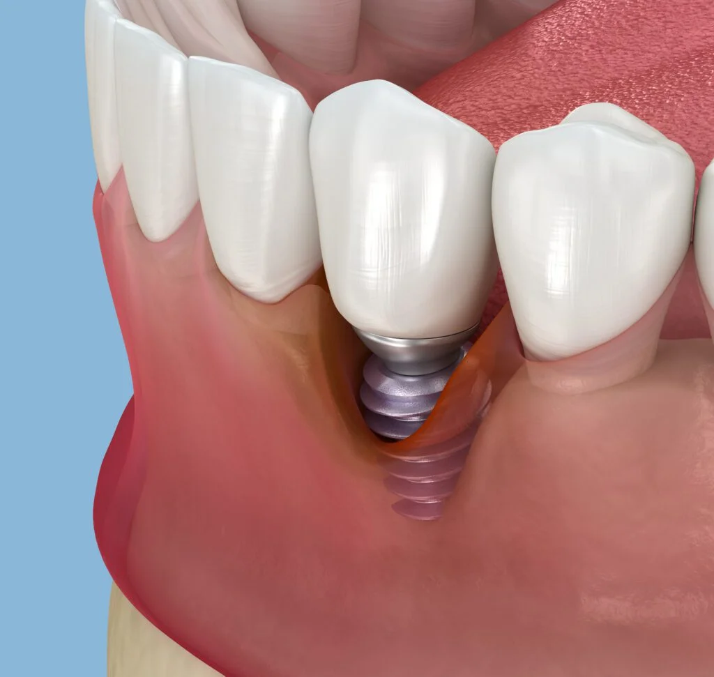
Peri-implant disease begins as peri-implant mucositis, a localized inflammation confined strictly to the soft gum tissue surrounding the implant collar. If left unmanaged, the bacterial colonies penetrate deeper along the implant threads, releasing toxins that trigger a destructive host immune response. This leads to peri-implantitis: a chronic destructive infection that leads to progressive loss of the supporting jawbone.
Recognizing Peri-Implantitis Symptoms and Early Warning Signs
Detecting early peri-implantitis allows for conservative, non-surgical intervention before irreversible bone damage occurs. Common peri-implantitis symptoms include:
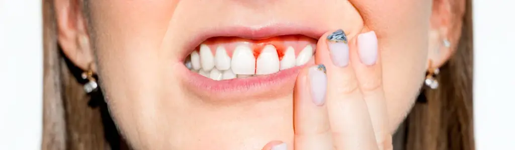
- Bleeding on Brushing or Probing: The earliest and most reliable clinical marker of active inflammation.
- Erythema and Edema: The gum tissue surrounding the implant appears bright red, puffy, or shiny instead of pale pink and firm.
- Deepening Pocket Depths: Routine periodontal probing shows pocket depths increasing from healthy measurements (2 to 3 mm) to 5 mm or deeper.
- Suppuration (Pus Discharge): Active infection produces small amounts of yellowish exudate or pus when the gum margin is lightly pressed.
- Exposed Implant Threads: As bone and gum tissue recede, the dark gray metallic threads of the implant fixture become visible through or above the gumline.
- Dull Aching or Throbbing: While early stages are often painless, advanced infection can cause localized tenderness during chewing.
- Implant Mobility: Movement of the implant fixture is a late-stage symptom indicating complete loss of osseointegration, meaning the implant cannot be saved.
The Clinical Peri-Implantitis Stages
Dental professionals categorize bone resorption into distinct peri-implantitis stages based on clinical probing depths and radiographic bone loss:
Stage 1: Initial / Early Peri-Implantitis
- Probing depths reach 4 to 5 mm.
- Minor horizontal or vertical bone loss (less than 25 percent of the implant length or less than 2 mm from initial placement).
- Bleeding on gentle probing with mild redness.
Stage 2: Moderate Peri-Implantitis
- Probing depths increase to 6 to 7 mm.
- Radiographs show saucer-shaped bone loss between 25 and 50 percent of the total implant length.
- Frequent suppuration, bleeding, and localized gum recession exposing 1 to 2 implant threads.
Stage 3: Advanced / Severe Peri-Implantitis
- Probing depths exceed 8 mm.
- Bone loss exceeds 50 percent of the total implant fixture length.
- Massive crater-like bone defects surrounding the fixture. At this stage, extraction and bone grafting are often necessary unless aggressive surgical decontamination is performed.
Critical Peri-Implantitis Risk Factors
Understanding the primary peri-implantitis risk factors helps patients prevent reinfection and identify personal vulnerabilities:
- Poor Oral Hygiene: Inadequate brushing, lack of water flossing, and missing 6-month professional cleanings allow bacterial plaque to build into calcified calculus on implant surfaces.
- History of Periodontitis: Patients who previously lost natural teeth to severe gum disease harbor periodontal pathogens that can infect dental implants.
- Tobacco and Nicotine Use: Smoking and vaping constrict micro-blood vessels, suppress local immune responses, and increase the risk of peri-implantitis by three to four times.
- Uncontrolled Diabetes Mellitus: Elevated blood sugar levels impair collagen synthesis, delay wound healing, and exacerbate bacterial inflammation around bone tissue.
- Excess Subgingival Cement: When dental crowns are cemented with traditional glue rather than screw-retained, excess cement pushed beneath the gumline acts as a rough foreign body that harbors bacteria.
- Occlusal Overload (Bruxism): Heavy clenching or grinding generates micro-fractures in the surrounding bone, creating pathways for bacterial invasion.
Modern Peri-Implantitis Treatment: Non-Surgical, Surgical, and Laser Protocols
A structured approach to peri-implantitis treatment aims to eliminate bacterial biofilm, decontaminate titanium surfaces, and regenerate lost bone structure.
TREATMENT HIERARCHY:
- Decontamination: Remove plaque & biofilm using titanium-friendly instruments.
- Laser Disinfection: Eliminate 99.9% of subgingival pathogens using Er:YAG / Nd:YAG.
- Surgical Debridement: Open flap access for deep decontamination of implant threads.
- Bone Regeneration: Place bone grafts & collagen membranes (guided bone regeneration).
1. Non-Surgical Mechanical Debridement
In early stages, specialized carbon fiber, plastic, or titanium curettes (which do not scratch the smooth implant collar) are used alongside glycine or erythritol air-polishing powder to strip away soft bacterial biofilms.
2. Advanced Laser Treatment of Peri-Implantitis Turkey
Medical tourism centers in Turkey are global leaders in minimally invasive laser treatment of peri-implantitis Turkey. Using advanced laser wavelengths (such as Er:YAG, CO2, and diode lasers), specialists can:
- Vaporize anaerobic bacteria from micro-porous titanium threads without overheating adjacent bone.
- Remove infected granulation tissue with minimal bleeding and zero mechanical scratching.
- Biostimulate surrounding bone cells (photobiomodulation) to promote natural tissue reattachment.
3. Surgical Decontamination and Implantoplasty
For moderate to severe cases, a periodontal surgeon opens a small gum flap to directly expose the affected threads. The exposed threads are mechanically smoothed (implantoplasty) using fine diamond burs to make the metal surface non-retentive to bacteria.
4. Guided Bone Regeneration (GBR)
Once the implant is sterile, the crater-like bone defect is filled with synthetic or bovine bone mineral grafts and covered with a resorbable collagen barrier membrane to regrow lost jawbone support.
Treatment Modalities Comparison Matrix
| Treatment Modality | Target Stage | Invasiveness | Bacterial Elimination | Bone Regrowth Potential | Average Recovery |
| Mechanical Air-Polishing | Mucositis / Early Stage 1 | Non-invasive | Moderate (Biofilm removal) | None (Stops loss only) | 1 to 2 Days |
| Laser Phototherapy (Er:YAG) | Stage 1 & Stage 2 | Minimally invasive | Extremely High (>99,9%) | Moderate (Stimulates healing) | 3 to 5 Days |
| Open Flap Implantoplasty | Stage 2 & Stage 3 | Surgical | High (Mechanical smoothing) | Low to Moderate | 7 to 10 Days |
| Regenerative Bone Grafting | Moderate Stage 2 / 3 | Surgical | High (Combined with flap) | High (Structural rebuild) | 10 to 14 Days |
Frequently Asked Questions (FAQ)
Can a loose dental implant be saved if I have peri-implantitis?
Unfortunately, no. Once a dental implant becomes physically loose (mobile), it means complete loss of osseointegration has occurred. A loose implant cannot re-fuse with the bone and must be removed. The site must be cleaned, grafted with new bone, and allowed to heal for 4 to 6 months before a new implant can be placed.
What is the difference between peri-implant mucositis and peri-implantitis?
Peri-implant mucositis is a superficial inflammation confined strictly to the soft gum tissue, with no bone loss; it is 100 percent reversible with professional cleaning. Peri-implantitis is a deeper, destructive infection that involves irreversible loss of the supporting jawbone surrounding the implant.
Is laser treatment for peri-implantitis painful?
No. Laser therapy is performed under routine local anesthesia and is virtually painless. Because dental lasers cauterize blood vessels and seal nerve endings as they work, patients experience significantly less post-operative swelling, bleeding, and discomfort compared to traditional open scalpels.
How do I prevent peri-implantitis after my dental implant surgery?
To prevent peri-implant infections, brush twice daily with a soft-bristled or electric toothbrush, clean beneath crowns and bridges daily using a water flosser or interdental brushes, avoid smoking, and visit your dental clinic every 6 months for professional implant maintenance.

 |
|