Skip to content
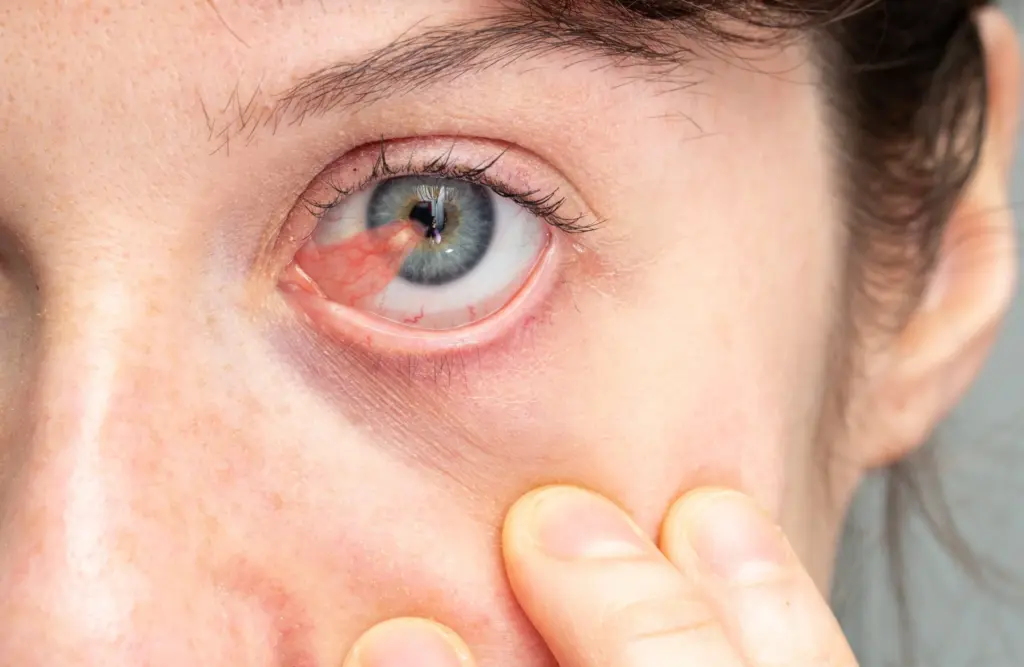
A pterygium, commonly known as “surfer’s eye”, is an elevated, wedge-shaped, non-cancerous fibrovascular growth that originates on the clear mucous membrane of the eye (the conjunctiva) and slowly extends across the limbus onto the cornea. Chronic exposure to ultraviolet (UV) radiation, environmental wind, fine sand, and chronic ocular surface dryness trigger this proliferative tissue response. While early lesions cause mild redness and foreign body sensations managed with lubricating drops, advancing lesions physically distort the corneal curvature, induce irregular astigmatism, cause significant visual impairment, and compromise facial aesthetics.
When non-surgical management fails or the growth approaches the central visual axis, pterygium surgery provides the definitive clinical intervention.
Table of Contents
What Is Pterygium Surgery and How Does It Work?
If you are wondering what is pterygium surgery, it is an ophthalmic microsurgical procedure designed to excise the fibrovascular lesion completely from the cornea and sclera, followed by ocular surface reconstruction to minimize the risk of recurrence.
Get Free Consultation
Have any questions? Leave your details and we'll get back to you shortly.
Historically, surgeons relied on the “bare sclera” technique, in which the growth was simply stripped away, leaving the underlying white of the eye exposed to re-epithelialize naturally. However, this outdated approach resulted in unacceptable recurrence rates between 40 and 80 percent, often producing a secondary growth more aggressive than the original lesion.
Modern ophthalmic surgery relies on the Conjunctival Autograft (CAG) technique, recognized globally as the gold standard:
CONJUNCTIVAL AUTOGRAFT SURGICAL SEQUENCE:
- [Corneal & Scleral Dissection]: Pterygium carefully peeled & excised under high-magnification microscope
- [Scleral Bed Smoothing]: Micro-burr polishes the corneal surface to restore optical clarity
- [Donor Tissue Harvest]: Healthy conjunctival tissue harvested from beneath the upper eyelid
- [Fibrin Tissue Glue Fixation]: Autograft secured over bare sclera using sutureless biological adhesive
By harvesting a thin patch of healthy, unexposed conjunctiva from under the patient’s upper eyelid and transplanting it onto the surgical site, limbal stem cells are restored. When secured with human fibrin tissue adhesive (glue) rather than traditional nylon sutures, recurrence rates drop to less than 2 to 5 percent while dramatically accelerating postoperative recovery.
Surgical Discomfort and Anesthesia: Is Pterygium Surgery Painful?
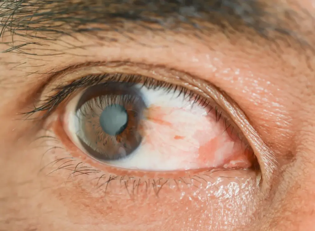
A primary concern among prospective ophthalmic patients is: is pterygium surgery painful?
- During Surgery: The procedure itself is completely painless. The ophthalmic surgeon administers topical anesthetic eye drops (such as proparacaine or tetracaine), frequently combined with a local subconjunctival anesthetic block. Patients remain awake, relaxed, and comfortable, feeling only light pressure, fluid irrigation, or cool water sensations throughout the 20- to 30-minute procedure.
- Early Postoperative Period: As local anesthesia subsides over the first 2 to 4 hours, patients experience mild to moderate ocular soreness. Common sensations include a gritty feeling (as if a grain of sand or an eyelash is trapped under the eyelid), eye watering, and light sensitivity. When sutureless biological fibrin glue is used instead of sutures, tissue friction against the inner eyelid is virtually eliminated, reducing postoperative pain scores by more than 70 percent. Oral analgesics and cold compresses easily manage early discomfort.
Visual & Aesthetic Expectations: Pterygium Surgery Before and After
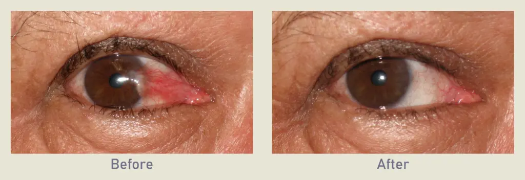
Reviewing documented clinical outcomes in pterygium surgery before and after cases demonstrates clear functional and visual rehabilitation:
- Corneal Smoothness and Astigmatism Correction: Advanced pterygia mechanically pull and flatten the corneal meridian, inducing irregular astigmatism. Surgical excision restores the normal spherical shape of the cornea, stabilizing refraction and improving uncorrected visual acuity.
- Aesthetic Restoration of the Sclera: The inflamed, fleshy, red triangular growth crossing the iris is removed. Once healed, the underlying sclera regains its clear, white appearance, blending seamlessly with the surrounding conjunctiva.
- Elimination of Chronic Ocular Irritation: Patients experience immediate relief from chronic burning, dryness, reflex tearing, and the persistent sensation of a foreign object in the eye.
Recovery Milestones: Pterygium Surgery Recovery Time
The pterygium surgery recovery time progresses across distinct anatomical healing phases:
- Days 1 to 3 (Acute Healing Phase): An eye patch or protective shield is worn over the operated eye for the first 24 hours. Once the patch is removed, mild redness, swelling (chemosis), and tearing are expected. Patients begin a strict regimen of antibiotic and topical corticosteroid eye drops to prevent infection and suppress tissue inflammation.
- Days 4 to 7 (Surface Re-Epithelialization): The superficial corneal epithelium heals and seals completely. Light sensitivity diminishes, and patients working sedentary or desk jobs can comfortably return to work within 3 to 5 days.
- Weeks 2 to 4 (Graft Integration): The conjunctival autograft firmly bonds to the underlying scleral vascular bed. The initial pinkish hue of the transplanted graft fades toward a clear, natural white. Driving and non-strenuous daily activities are resumed.
- Months 2 to 3 (Final Stabilization): Topical steroid drops are gradually tapered under slit-lamp microscopic supervision. The corneal clarity stabilizes permanently.
Guidelines for Care After Pterygium Surgery
Proper self-care after pterygium surgery and following your post pterygium surgery protocols prevents surgical complications and tissue relapse:
- Adhere to the Prescribed Eye Drop Protocol: Administer broad-spectrum antibiotic and anti-inflammatory steroid eye drops exactly as scheduled to prevent graft edema and fibrosis.
- Never Rub the Operated Eye: Avoid rubbing or pressing on the eye for at least 4 to 6 weeks, as mechanical friction can dislodge or shear the delicate autograft before vascular anchoring is complete.
- Wear 100% UV-Blocking Sunglasses: Direct sunlight is the primary environmental driver of pterygium development and recurrence. Wrap-around sunglasses with UV400 protection must be worn whenever outdoors.
- Avoid Water and Contaminated Environments: Keep tap water, soap, and shampoo away from the eye for the first 7 to 10 days. Swimming pools, hot tubs, dusty worksites, and sandy beaches must be avoided for at least one month.
Understanding Potential Pterygium Surgery Side Effects
While modern sutureless autografting is a safe outpatient surgery, patients should understand potential pterygium surgery side effects:
- Graft Displacement or Loss: Accidental eye rubbing in the early postoperative days can displace the autograft, occasionally requiring surgical re-positioning.
- Prolonged Subconjunctival Redness: The graft area remains pink or mildly injected for several weeks due to active capillary healing before blanching out completely.
- Corneal Dellen: A localized thinning or temporary dry indentation on the corneal surface adjacent to the surgical margin, easily treated with frequent preservative-free artificial tears and temporary eye patching.
- Pyogenic Granuloma: A benign, vascularized, fleshy inflammatory nodule that can form along the graft incision line, typically resolving with topical steroid adjustments or minor in-office excision.
- Pterygium Recurrence: When modern autografting with fibrin glue is performed, the risk of recurrence is minimized to less than 5 percent, compared to over 50 percent with older excision methods.
Comparison of Surgical Techniques for Pterygium Removal
| Surgical Technique | Recurrence Rate | Postoperative Discomfort | Procedure Time | Graft Anchoring Method | Overall Safety & Efficacy |
| Bare Sclera Excision | High (40% to 80%) | Moderate | 10 to 15 Minutes | None (Sclera left open) | Outdated; Not Recommended |
| Autograft with Sutures | Low (5% to 10%) | Moderate to High (Foreign body feel) | 35 to 45 Minutes | Absorbable / Nylon Sutures | Reliable; Higher tissue irritation |
| Sutureless Autograft (Fibrin Glue) | Extremely Low (<2% to 5%) | Minimal | 20 to 30 Minutes | Biological Fibrin Adhesive | Gold Standard Modern Method |
| Amniotic Membrane Graft | Low to Moderate (10% to 15%) | Minimal to Moderate | 25 to 35 Minutes | Fibrin Glue or Fine Sutures | Reserved for large/recurrent cases |
What Factors Determine the Pterygium Surgery Cost?
When evaluating pterygium surgery cost, patients will find that pricing is variable rather than a standardized flat fee. The total expense is shaped by several distinct clinical, geographic, and facility variables:
- Surgical Technique and Grafting Modality: Modern sutureless conjunctival autografting using medical-grade biological fibrin adhesive carries higher material expenses than basic suture techniques, but delivers lower recurrence rates and higher patient comfort.
- Unilateral vs. Bilateral Procedures: Treating a single eye involves a different operational scope and theater time than simultaneous bilateral surgery or large, double-headed pterygia (growths originating from both the nasal and temporal sides of the same eye).
- Primary vs. Recurrent (Revision) Surgery: Removing a recurrent pterygium that has scarred down from a previous failed operation is technically demanding, frequently requiring amniotic membrane transplantation or adjunctive antimetabolite medications (such as Mitomycin C), which adjust the surgical fee.
- Surgeon Expertise and Clinical Infrastructure: Corneal sub-specialists and anterior segment ophthalmic surgeons operating in fully accredited ophthalmic surgical suites with high-magnification surgical operating microscopes reflect their specialized training in their professional fees.
- Geographic Location and Package Inclusions: Healthcare systems in Western Europe and the US carry higher clinic overhead than medical tourism hubs such as Turkey. International patient packages in Turkey frequently bundle pre-op corneal topography scans, surgeon fees, surgical facility charges, post-op medications, protective eye shields, hotel stays, and private airport transfers into a single structured arrangement.
Frequently Asked Questions (FAQ)
Can a pterygium grow back after surgery?
While modern conjunctival autografting using fibrin glue has lowered recurrence rates to under 5 percent, a pterygium can theoretically return if the eye is exposed to heavy ultraviolet (UV) light, wind, dust, and persistent dryness during the recovery period. Wearing wrap-around UV-blocking sunglasses and keeping the ocular surface lubricated with preservative-free artificial tears is essential for long-term prevention.
How soon after pterygium surgery can I drive and return to work?
Most patients can drive safely within 48 to 72 hours once the eye patch is removed and visual acuity clears. Patients working desk-based jobs or remote computer work typically return within 3 to 5 days, while those working in dusty, outdoor, or physically demanding environments should wait 10 to 14 days to prevent contamination.
Is it better to use fibrin glue or sutures for pterygium autografts?
Fibrin tissue adhesive (glue) is superior to traditional sutures for the majority of patients. Clinical trials demonstrate that fibrin glue significantly shortens surgical duration, reduces postoperative pain and foreign body sensation, prevents suture-related tissue inflammation, and produces a lower recurrence rate compared to nylon or Vicryl sutures.
When should I consider having pterygium surgery rather than waiting?
Surgery is indicated when the pterygium causes chronic redness, burning, or discomfort unresponsive to lubricating eye drops; induces progressive corneal astigmatism and blurred vision; extends toward the pupillary margin threatening the central visual axis; or causes significant cosmetic self-consciousness.

 |
|