Skip to content

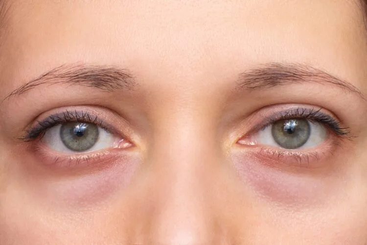
The periorbital zone is among the most structurally complex areas of facial anatomy. Because the cutaneous layer beneath the eyes is the thinnest in the human body, any alteration in volume, fat cushion stability, or vascular flow becomes quickly noticeable. Swelling, puffiness, and protruding contours across the lower eyelid, collectively termed eye bags, frequently convey an impression of chronic exhaustion, illness, or accelerated facial aging. While mild morning puffiness often responds to simple lifestyle modifications, genuine structural lower eyelid bags involve deep anatomical shifts that cannot be corrected by eye creams or topical cold rollers alone.
Individuals seeking to rejuvenate their periorbital contour routinely investigate what causes eye bags, seek to differentiate between distinct types of eye bags, look for actionable protocols on how to get rid of eye bags, analyze biological factors underlying eye bags causes, explore clinical options for an under eye bags treatment, evaluate surgical steps on how to remove eye bags permanently, and assess overall investments for a definitive eye bags treatment. This clinical guide examines orbital biomechanics, anatomical classifications, surgical and non-surgical procedures, recovery benchmarks, and cost variables.
Table of Contents
Anatomical Classifications: Understanding the Types of Eye Bags
Effective clinical correction begins with a precise diagnosis. In oculoplastic surgery, patients are evaluated based on several distinct types of eye bags:
Get Free Consultation
Have any questions? Leave your details and we'll get back to you shortly.
- Pseudoherniated Orbital Fat Bags: The most prevalent structural presentation. Intraorbital fat cushions (divided into medial, central, and lateral compartments) naturally protect the eyeball. When the fibrous retaining wall, the orbital septum, weakens due to genetics or aging, these fat pads bulge outward, producing persistent, soft, rounded protrusions under the lower eyelid margin.
- Fluid Retention and Edematous Bags (Malar Mounds): Caused by impaired microvascular or lymphatic drainage within the periorbital tissue. These bags fluctuate dramatically throughout the day, presenting with severe swelling upon waking that gradually dissipates as gravity assists lymphatic clearing during upright hours.
- Cutaneous and Muscular Laxity Bags (Dermatochalasis): Characterized by excessive stretching of the lower eyelid skin alongside laxity of the orbicularis oculi muscle. Rather than firm fat protrusions, these bags present as crepey, accordion-like folds of loose, wrinkled skin that gather beneath the eyelashes.
- Festoons and Malar Bags: Complex tissue swellings positioned lower on the face, resting over the zygomatic bone (upper cheek). They involve chronic inflammation of the sub-orbicularis oculi fat (SOOF), localized lymphedema, and attenuation of the orbito-malar retaining ligament.
- Structural Hollow Bags (Negative Vector Anatomy): In individuals with genetic midface hypoplasia or deficient cheekbones, the eyeball rests anterior to the lower orbital rim. This structural depression under the eye creates a harsh shadow that mimics a protruding eye bag.
Pathophysiological Triggers: What Causes Eye Bags?
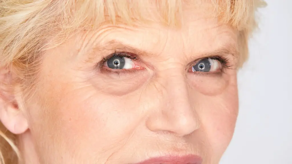
Investigating what causes eye bags and analyzing biological eye bags causes reveals a convergence of genetic, structural, and environmental factors:
- Attenuation of the Orbital Septum: Over time, dermal collagen synthesis declines and elastolytic degradation accelerates. The orbital septum loses its tensile strength, allowing intraorbital fat to herniate forward under normal ocular pressure.
- Maxillary Bone Resorption: With advancing age, the underlying craniofacial skeletal foundation undergoes natural bone loss. The lower orbital rim recedes backward, reducing the physical shelf that supports the lower eyelid structures and leaving space for tissues to fall forward.
- High-Sodium Diet and Fluid Shifts: Consuming high-sodium meals close to bedtime prompts osmotic fluid retention in interstitial tissues. The loose areolar connective tissue beneath the eyes readily absorbs excess fluid, manifesting as severe morning puffiness.
- Allergic Rhinitis and Histamine Release: Environmental allergies provoke localized mast cell degranulation, triggering histamine-induced capillary permeability. Chronic venous congestion in the periorbital venous plexus compounds both edema and dark circles.
- Chronic Sleep Deprivation: Poor sleep disrupts systemic cortisol rhythms and impairs cutaneous microcirculation, leading to vascular dilation and poor lymphatic clearance.
- Genetic Heredity: Many young individuals in their teens and twenties exhibit prominent pseudoherniated fat pads solely due to inherited familial anatomical traits, occurring independently of lifestyle habits.
Non-Surgical Modalities: Under Eye Bags Treatment
For patients presenting with mild fluid accumulation, slight volume loss, or early skin laxity, non-invasive under eye bags treatment options offer temporary aesthetic refinement:
1. Hyaluronic Acid Dermal Fillers (Tear Trough Camouflage)
When minor orbital fat herniation is accentuated by a deep adjacent tear trough groove, strategic injection of low-viscosity, non-hydrophilic hyaluronic acid gel can smooth the transition. Using a blunt microcannula, the filler is deposited in the deep pre-periosteal plane along the orbital rim. By elevating the hollow shadow beneath the bag, the protrusion is visually camouflaged.
2. Fractional Laser Resurfacing (CO2 and Erbium Lasers)
Indicated for fine lines, crepey skin texture, and mild cutaneous laxity. The laser creates thousands of microscopic thermal ablation zones in the lower eyelid dermis. This controlled thermal injury triggers wound repair, stimulating healthy collagen contraction and tightening loose surface skin.
3. Radiofrequency (RF) Microneedling and Plasma Sublimation
RF microneedling delivers focused thermal energy directly into the subdermal layers, coagulating loose collagen fibers and thickening the delicate skin barrier without damaging surface pigmentation.
4. Topical Formulations and Lymphatic Drainage
Serums containing pharmaceutical-grade caffeine, peptide complexes (such as acetyl tetrapeptide-five), and vitamin K temporarily constrict superficial blood vessels and stimulate microvascular fluid clearance, easing mild morning puffiness.
Definitive Surgical Solutions: How to Remove Eye Bags Permanently?
When addressing pseudoherniated fat pads, marked structural bulging, or severe tissue excess, patients often ask how to remove eye bags permanently. Surgical blepharoplasty remains the clinical gold standard:
1. Transconjunctival Lower Blepharoplasty (Scarless Approach)
The preferred technique for younger patients and individuals with protruding fat pads but good skin elasticity.
- Incision Placement: The plastic surgeon makes a delicate horizontal incision along the mucosal inside of the lower eyelid (the palpebral conjunctiva).
- Fat Redraping or Excision: Through this internal window, the surgeon accesses the medial, central, and lateral fat compartments. Rather than aggressively cutting out all the fat, which can cause an aged, hollowed appearance, modern techniques preserve and reposition the fat over the lower orbital rim (fat redraping) to fill deep tear troughs permanently.
- Zero External Scar: Because the incision is entirely internal, no visible cut or scar exists on the external skin, and the structural shape of the eye aperture remains unaltered.
2. Transcutaneous Lower Blepharoplasty (Subciliary Approach)
Indicated for mature patients who present with prominent fat herniation accompanied by severe, redundant skin folds and muscle laxity.
- Incision Placement: The incision runs externally along the lower lash line, hidden within a natural crease.
- Tissue Tailoring: The surgeon trims protruding fat pads, lifts and tightens the lax orbicularis oculi muscle (canthopexy or canthoplasty to reinforce lateral support), and excises a measured strip of excess skin before closing with fine micro-sutures.
Recovery Roadmap: What to Expect During Post-Operative Healing
Following surgical correction, understanding the healing trajectory ensures a safe, smooth recovery:
- Days One to Three (Acute Inflammatory Phase): Swelling and bruising around the lower eyelids and upper cheeks peak around day three. Patients apply cold gel compresses for fifteen minutes every waking hour. Tightness, mild blurred vision from lubricating eye ointments, and light sensitivity are normal.
- Days Four to Seven (Early Stabilization): External subciliary sutures are removed on day five to seven. Internal transconjunctival incisions heal autonomously without suture removal. Bruising transitions from purple to light yellowish-green.
- Weeks Two to Three (Social Recovery): Most residual swelling and discoloration resolve. Patients comfortably return to sedentary work, public social settings, and light aily walking. Makeup can usually be resumed once skin incisions are fully sealed.
- Months One to Six (Final Maturation): Deep tissue remodeling completes. The lower eyelid contour settles into a flat, youthful transition that flows continuously into the upper cheek.
Visual Transformations: Clinical Before and After Expectations
Documented clinical cases illustrate the substantial functional and cosmetic improvements achieved through targeted treatment:
- Elimination of Protruding Fat Bulges: Herniated fat pads are removed or flattened, restoring a smooth, flat lower eyelid platform.
- Eradication of Deep Shadow Lines: Redraping fat into the tear trough erases the harsh optical shadow separating the lower lid from the midface.
- Smoothing of Wrinkled, Crepey Skin: Excision of loose cutaneous tissue eliminates accordion-like folds, providing a tighter surface under makeup.
- Awakened, Refreshed Expression: The unintentional look of chronic fatigue, aging, or exhaustion is replaced by a vibrant, rested periorbital appearance.
Essential Practical Guidelines: How to Get Rid of Eye Bags
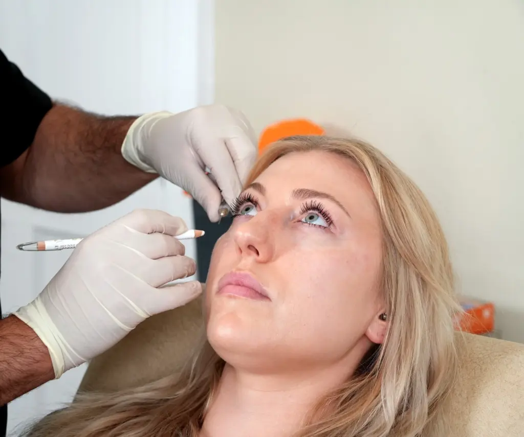
Whether recovering from clinical interventions or minimizing aily fluid retention, follow these guidelines to manage periorbital swelling:
- Keep the Head Elevated at Night: Sleep with your head elevated on two pillows or an ergonomic wedge. Elevating the head above heart level harnesses gravity to prevent overnight venous and lymphatic pooling.
- Eliminate High-Sodium Dinners: Restrict sodium consumption during evening meals to reduce fluid shift into the loose periorbital tissues.
- Apply Cold Compresses Correctly: Use clean, cold gel packs wrapped in a soft cloth. Never place raw ice directly on the delicate lower eyelid skin, as thermal freezing can injure fragile surface capillaries.
- Avoid Eye Rubbing: Chronic friction stretches the thin lower lid skin, degrades structural collagen, breaks fragile capillaries, and worsens pigmentary dark circles.
- Wear UV-Blocking Sunglasses: Ultraviolet radiation accelerates elastosis and degrades the orbital septum. Wearing broad-spectrum UV400 sunglasses protects structural tissues and prevents repetitive squinting.
Comparative Matrix: Eye Bag Interventions
| Clinical Modality | Topical Serums & Compresses | Tear Trough Dermal Fillers | Fractional Laser Resurfacing | Lower Blepharoplasty Surgery |
| Primary Indication | Mild morning fluid retention | Structural tear trough shadows | Crepey, loose surface skin | Herniated fat pads & severe bags |
| Mechanism | Microvascular constriction | Hyaluronic acid volume filling | Thermal dermal neocollagenesis | Surgical fat excision / redraping |
| Invasiveness | Completely non-invasive | Minimally invasive (cannula) | Non-surgical thermal ablation | Outpatient surgical intervention |
| Procedural Duration | Daily home application | 20 to 30 Minutes | 20 to 45 Minutes | 60 to 90 Minutes |
| Discomfort Level | None | Mild (topical anesthetic) | Moderate (topical numbing) | Mild (local with IV sedation) |
| Downtime | Zero downtime | 2 to 4 Days (mild bruising) | 5 to 7 Days (peeling & redness) | 10 to 14 Days (social recovery) |
| Longevity of Results | Temporary (hours) | 9 to 18 Months | 1 to 2 Years | Permanent (10 to 20 years) |
What Determines the Eye Bags Treatment Cost?
Prospective patients researching the overall financial commitment for periorbital rejuvenation will find that costs vary widely based on clinical, technological, and anatomical parameters:
- Complexity of the Surgical Approach: A scarless transconjunctival blepharoplasty involves different surgical equipment and operative time than an open subciliary approach that combines fat repositioning, lateral canthopexy, and skin excision.
- Need for Ancillary Procedures: Many patients require combined interventions to address multiple aging mechanisms simultaneously, such as adding fat grafting (nanofat) to correct hollow cheeks, or fractional laser resurfacing to treat sun-damaged skin.
- Anesthesia and Facility Standards: Treatments performed under general anesthesia or monitored intravenous sedation in an accredited hospital surgical suite carry different operational facility costs than non-surgical injections done in an outpatient clinic room.
- Surgeon Specialization and Board Certification: Highly experienced oculoplastic surgeons, certified facial plastic surgeons, and general plastic surgeons with dedicated fellowship training in periorbital reconstructive surgery structure fees reflecting their clinical precision and low revision rates.
- Geographic Destination and All-Inclusive Healthcare Packages: Operational overhead, malpractice structures, and currency dynamics create marked pricing variations between private clinics in North America or Western Europe and internationally accredited healthcare hubs like Turkey. Comprehensive aesthetic travel packages in Turkey frequently bundle pre-operative diagnostic evaluations, surgical hospital fees, medications, boutique hotel lodging for six to eight nights, and private VIP transfers into structured care arrangements.
Frequently Asked Questions (FAQ)
Can eye creams completely eliminate hereditary eye bags?
No. Topical eye creams, caffeine serums, and moisturizers can hydrate the epidermis, temporarily tighten surface crepeyness, and slightly ease morning fluid accumulation. However, they cannot penetrate through the skin, muscle, and fibrous orbital septum to dissolve or shrink deep herniated fat pads. True structural fat bags require clinical or surgical intervention.
Do eye bags come back after lower blepharoplasty surgery?
In the vast majority of cases, the results of a lower blepharoplasty are permanent and endure for decades. Once herniated intraorbital fat cells are excised or repositioned, they do not regrow. While natural facial aging and tissue thinning will continue over time, recurring fat herniation requiring a secondary revision is exceedingly rare.
Can under-eye dermal fillers make eye bags look worse?
Yes, if injected improperly. Placing a hydrophilic hyaluronic acid filler directly over a herniated fat pad or superficially within the lymphatic plane can absorb excess water, worsening the appearance of the bag and producing a bluish hue known as the Tyndall effect. Fillers should only be placed in the deep pre-periosteal hollow beneath the bag by an experienced injector.
What is the difference between eye bags and festoons?
True eye bags are localized within the lower eyelid margin directly beneath the eyelashes, caused by herniated orbital fat or loose eyelid skin. Festoons (malar bags) are situated lower on the face, resting over the cheekbone (malar eminence). Festoons involve chronic lymphatic swelling, sun-damaged muscle laxity, and fluid accumulation within the cheek tissues, requiring specialized skin resurfacing or extended surgical suspension rather than standard lower blepharoplasty alone.

 |