Skip to content
Intraocular lens (IOL) implantation is one of the most performed and successful ophthalmic procedures worldwide. During standard cataract surgery or refractive lens exchange, an eye surgeon removes the cloudy or defective natural lens and replaces it with an artificial acrylic or silicone intraocular lens. In the vast majority of cases, the artificial lens rests securely inside the natural capsular bag behind the pupil, providing crisp, clear vision for life.
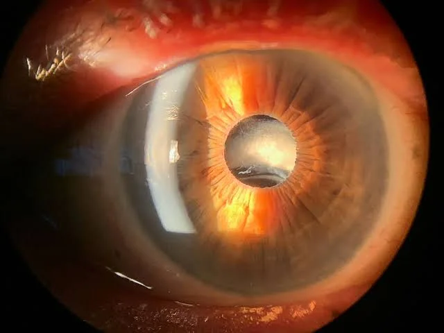
However, years after an otherwise flawless surgery or following physical trauma, the internal supportive structure of the eye can weaken. This weakening can cause the implant to shift out of alignment, leading to a dislocated intraocular lens.
If you or a loved one suspect an implant has shifted, understanding the condition is vital for preserving vision. Patients frequently search for the primary signs of dislocated intraocular lens, examine visual aids like a dislocated intraocular lens diagram, monitor specific dislocated intraocular lens symptoms, and seek information regarding surgical treatment for dislocated intraocular lens. This comprehensive clinical guide explores why a dislocated intraocular lens (IOL) occurs, details diagnostic steps, and outlines modern surgical repositioning and exchange procedures.
Get Free Consultation
Have any questions? Leave your details and we'll get back to you shortly.
Table of Contents
What Is a Dislocated Intraocular Lens (IOL)?
To understand how an implant shifts, we must examine the internal anatomy of the eye. During primary cataract surgery, the surgeon creates a circular opening in the front of the natural lens capsule (capsulorhexis), clears out the cataract, and places the artificial lens inside the remaining clear capsule. This natural envelope is suspended in place behind the iris by microscopic, hair-like fibers called zonules (zonules of Zinn).
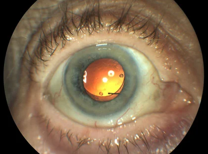
A dislocated intraocular lens occurs when these supportive zonular fibers stretch, snap, or break away from the eye wall. When a few zonules break, the lens tilts or shifts off-center (subluxation). If all or most zonular attachments fail, the entire lens can fall backward into the vitreous cavity at the back of the eye (complete dislocation).
Primary Causes: Why Does an Intraocular Lens Dislocate?
An artificial lens rarely fails on its own; rather, dislocation occurs because the underlying ocular tissues holding the capsular bag lose structural integrity. Key triggers include:
- Pseudoexfoliation Syndrome (PXF): A systemic metabolic condition where abnormal protein fibrillar material deposits on the lens capsule and zonules, causing them to become extremely brittle and break over time.
- Blunt Ocular Trauma: A direct physical blow to the eye, head, or face from sports injuries, falls, or accidents can immediately snap fragile zonular fibers.
- Prior Vitreoretinal Surgery: Previous intraocular operations, particularly vitrectomy procedures, can stress and weaken zonular support.
- High Myopia (Severe Nearsightedness): Highly myopic eyes have an elongated eyeball structure, which naturally stretches and thins the zonular apparatus.
- Late Capsular Bag Contraction: Years after surgery, normal scar tissue inside the capsule can shrink, exerting uneven tension on weak zonules and pulling the lens off-center.
Recognizing the Warning Signs: Dislocated Intraocular Lens Symptoms
When an artificial lens moves out of its optical axis, light rays entering the eye no longer focus cleanly onto the retina. Identifying early signs of dislocated intraocular lens shifts helps prevent severe vision impairment. Key dislocated intraocular lens symptoms include:
- Progressive or Sudden Blurry Vision: Objects appear out of focus or distorted as the optical power of the lens moves away from the pupil center.
- Monocular Diplopia (Double Vision in One Eye): When the edge of the dislocated lens sits directly across the pupil, light enters through both the artificial lens and the open space beside it, creating a duplicate or shadowed image in that eye alone.
- Crescent-Shaped Shadow (Edge Effect): Patients often report seeing a dark, crescent-like shadow or ring at the edge of their peripheral visual field.
- Fluctuating Vision with Head Movement: If the lens is loosely tethered, tilt and clarity can change depending on whether you lean forward, lie flat, or turn your head.
- Visible Lens Edge in the Pupil: A doctor or family member may notice a glint, crescent, or visible plastic margin crossing the black center of the pupil.
- Aching Eye Pain and Redness: If a subluxated lens rubs against the iris or blocks internal fluid drainage, intraocular pressure can rise sharply, triggering deep aching pain and nausea.
Diagnostic Evaluation: How Ophthalmic Surgeons Confirm Dislocation
Confirming the position of a dislocated implant requires a detailed clinical exam by an ophthalmologist or vitreoretinal specialist:
- Slit-Lamp Biomicroscopy: Using a high-magnification microscope, the doctor evaluates the pupil after dilation. They check for lens tilt, visible zonular rupture, or the presence of a dislocated intraocular lens diagram-like crescent within the pupil space.
- Dilated Fundus Exam: Examining the back of the eye determines whether the lens has fallen completely into the posterior vitreous cavity.
- Anterior Segment Optical Coherence Tomography (AS-OCT): High-resolution 3D cross-sectional imaging provides precise measurements of lens tilt, vault, and distance from the corneal endothelium.
- B-Scan Ophthalmic Ultrasound: If dense vitreous hemorrhage or inflammation blocks the view, ultrasound waves map the location of a lens resting on the retina.
Surgical Solutions: Treatment for Dislocated Intraocular Lens
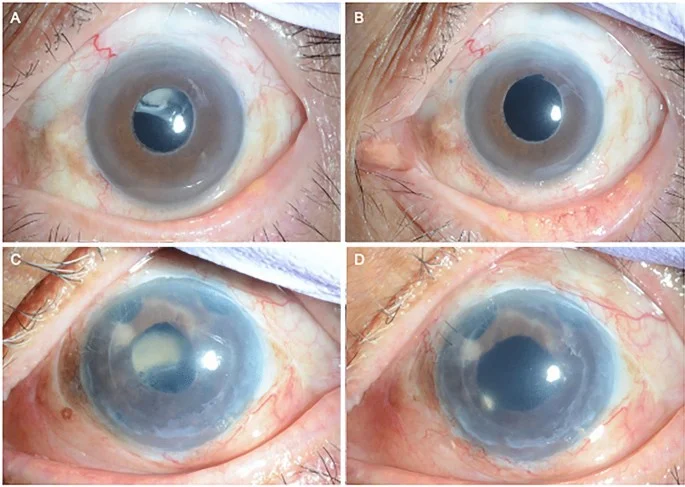
When an implant shifts enough to cause visual distortion or high pressure, conservative monitoring is no longer sufficient. Surgical treatment for dislocated intraocular lens requires specialized technique, often performed in tandem by an anterior segment surgeon and a vitreoretinal specialist.
1. Intraocular Lens Repositioning (Preserving the Original Lens)
If the dislocated lens is in good physical condition and matches the patient’s prescription, surgeons prefer to reposition and secure the existing implant:
- Scleral Fixation (Gore-Tex or Yamane Technique): The surgeon uses microscopic sutures or small terminal bulbs (flanges) created with a heated cautery tool to anchor the existing lens haptics directly into the white wall of the eye (sclera).
- Iris Suture Fixation: The haptics of the shifting lens are stitched directly to the back surface of the iris using micro-sutures.
2. Lens Retrieval and Exchange (Replacing the Shifted Implant)
If the existing lens is damaged, warped, or resting on the delicate retina at the bottom of the eye:
- Pars Plana Vitrectomy (PPV) with Lens Extraction: A retinal specialist uses specialized microsurgical tools to perform a vitrectomy, carefully lift the fallen lens off the retina, cut it into pieces if necessary, and remove it from the eye.
- Secondary IOL Implantation: The surgeon inserts a new artificial lens specifically designed for eyes without capsular support. Options include a scleral-fixated IOL, an anterior chamber IOL (placed in front of the iris), or an iris-claw lens (Yamane technique).
Comprehensive Surgical & Clinical Management Matrix
| Metric / Clinical Parameter | Mild Subluxation (Minor Tilt) | Moderate Subluxation (Partial Shift) | Complete Posterior Dislocation |
| Visual Impairment | Mild blur, slight glare | Double vision (monocular), crescent shadow | Severe drop in vision, aphakic state |
| Capsular Bag Condition | Intact with minor zonular strain | Partial zonular rupture (>180°) | Complete zonular collapse |
| Primary Treatment Approach | Observation / Corrective glasses | Surgical repositioning (Scleral fixation) | Pars Plana Vitrectomy + Lens exchange |
| Surgical Complexity | Non-surgical | Moderate (Anterior segment surgery) | High (Vitreoretinal team required) |
| Recovery Window | Immediate | 2 to 4 Weeks | 4 to 8 Weeks |
Frequently Asked Questions (FAQ)
Can a dislocated intraocular lens cause permanent blindness?
A dislocated intraocular lens does not automatically cause permanent blindness, but left untreated, severe cases can lead to vision-threatening complications. If a shifting lens causes sustained high intraocular pressure (secondary glaucoma), corneal damage, or retinal tears, permanent visual loss can occur. Prompt evaluation by an eye surgeon ensures a high probability of full visual recovery.
How quickly should surgery be performed for a dislocated intraocular lens?
The urgency of treatment depends on your symptoms and the location of the lens. If the lens has completely fallen onto the retina, is causing acute high eye pressure, or triggers severe pain, surgery is needed within days. If the lens is only mildly shifted and your vision is functional with glasses, surgery can be planned routinely over several weeks.
What is the Yamane technique for a dislocated intraocular lens?
The Yamane technique (scleral tunnel fixation) is an advanced, sutureless surgical method used to fixate a dislocated or secondary intraocular lens. The surgeon passes the flexible arms (haptics) of the lens through thin needles inserted into the white of the eye (sclera). Heat is applied to the end of each haptic to create a smooth, mushroom-shaped bulb (flange) that sits flush beneath the conjunctiva, anchoring the lens firmly in place without stitches.
What should I avoid doing if I suspect my intraocular lens has shifted?
If you suspect your lens has dislocated, avoid rubbing your eyes vigorously, refrain from heavy lifting or straining, and avoid sudden, violent head movements. Contact your ophthalmologist immediately for a dilated exam, and seek emergency care if you experience sharp eye pain, sudden flashes of light, or a curtain-like shadow in your vision.

 |
|